 뒤로
The Muscular System: Structure, Function, and Major Muscle Groups
스터디 가이드 - 스마트 노트
 자료에 맞춘 맞춤형 노트, 핵심 정의, 예시, 맥락을 확장해 제공합니다.
The Muscular System
Introduction to the Muscular System
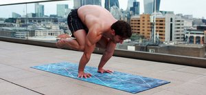
The muscular system is responsible for producing movement, maintaining posture, and generating heat in the human body. It consists of skeletal muscles, which are under voluntary control, and is essential for all forms of physical activity, from simple movements to complex athletic performances.
Movement: Muscles contract to move bones at joints, allowing for locomotion and manipulation of the environment.
Posture: Continuous muscle contractions help maintain body posture and position.
Heat Production: Muscle activity generates heat, contributing to the maintenance of body temperature.
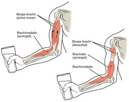
Muscle Actions: Prime Movers and Synergists
Muscle actions are coordinated by different types of muscles working together. The prime mover (agonist) is the main muscle responsible for a specific movement, while synergists assist the prime mover by adding force or reducing unnecessary movement.
Prime Mover (Agonist): The muscle primarily responsible for movement (e.g., biceps brachii in elbow flexion).
Synergist: Muscles that assist the prime mover (e.g., brachialis and brachioradialis in elbow flexion).
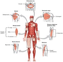
Muscle Shapes and Fiber Alignment
Skeletal muscles are classified based on their shapes and the arrangement of their fibers. These structural differences influence the muscle's function and strength.
Parallel: Fibers run parallel to the long axis (e.g., sartorius).
Pennate: Fibers attach obliquely to a central tendon (e.g., rectus femoris).
Convergent: Fibers converge from a broad origin to a single tendon (e.g., pectoralis major).
Circular: Fibers arranged in concentric rings (e.g., orbicularis oris).
Multipennate, Bipennate, Fusiform: Other arrangements that affect force and range of motion.
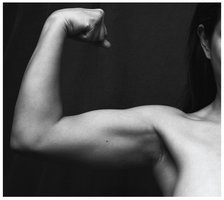
Muscle Structure: Belly and Tendons
The belly of a muscle is its thick, central part, while tendons are connective tissues that attach muscle to bone. Muscle contraction pulls on the tendons, moving the skeleton.
Belly: The central, contractile part of the muscle.
Tendons: Dense connective tissue that transmits the force of muscle contraction to bones.
Overview of the Muscular System: Superficial and Deep Muscles
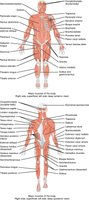
The human body contains both superficial (surface) and deep (underlying) muscles. Superficial muscles are visible just under the skin, while deep muscles lie beneath them and often stabilize joints or assist in complex movements.
Superficial Muscles: Responsible for gross movements and visible contours of the body.
Deep Muscles: Stabilize joints and support posture.
Understanding Muscle Names: Latin Roots
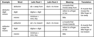
Muscle names often describe their location, action, shape, or size, and are derived from Latin roots. Understanding these roots helps in identifying muscle functions and positions.
Example | Word | Latin Root 1 | Latin Root 2 | Meaning | Translation |
|---|---|---|---|---|---|
abductor digiti minimi | abductor | ab = away from | duct = to move | a muscle that moves away from | A muscle that moves the little finger or toe away |
digiti | digitus = digit | refers to a finger or toe | |||
minimi | minimus = min, tiny | little | |||
adductor digiti minimi | adductor | ad = to, toward | duct = to move | a muscle that moves towards | A muscle that moves the little finger or toe toward |
digiti | digitus = digit | refers to a finger or toe | |||
minimi | minimus = min, tiny | little |
Major Muscle Groups and Their Functions
Muscles of Facial Expression
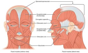
Facial muscles are unique because many insert into the skin rather than bones, allowing for a wide range of facial expressions. These muscles control movements of the eyelids, nose, and mouth.
Examples: Orbicularis oculi (closes eyelids), orbicularis oris (puckers lips), buccinator (compresses cheek).
Muscles of the Eyes
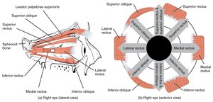
The extrinsic eye muscles originate on the skull and insert onto the eyeball, allowing for precise movements of the eye in different directions.
Examples: Superior rectus, inferior rectus, lateral rectus, medial rectus, superior oblique, inferior oblique.
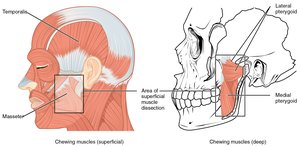
Muscles That Move the Lower Jaw (Mastication)
The muscles of mastication are responsible for moving the lower jaw during chewing. They originate from the skull and insert into the mandible, providing the leverage needed for biting and grinding food.
Examples: Masseter, temporalis, medial pterygoid, lateral pterygoid.
Muscles That Move the Tongue
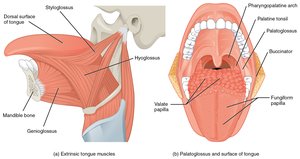
The tongue is moved by both intrinsic and extrinsic muscles, which are essential for speech, swallowing, and manipulating food.
Extrinsic Muscles: Genioglossus, hyoglossus, styloglossus.
Intrinsic Muscles: Change the shape of the tongue.
Muscles of the Anterior Neck
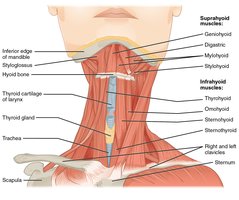
The anterior neck muscles facilitate swallowing and speech. They are divided into suprahyoid (above the hyoid bone) and infrahyoid (below the hyoid bone) groups.
Suprahyoid Muscles: Digastric, mylohyoid, stylohyoid, geniohyoid.
Infrahyoid Muscles: Sternohyoid, omohyoid, sternothyroid, thyrohyoid.
Muscles of the Neck and Back
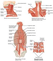
The neck and back muscles are responsible for moving the head, cervical vertebrae, and scapulas. They include both superficial and deep muscle layers.
Examples: Trapezius, splenius capitis, levator scapulae, semispinalis capitis.
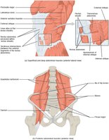
Muscles of the Abdomen
The abdominal wall is composed of several layers of muscles that protect internal organs, assist in breathing, and help with movements such as bending and twisting.
Rectus abdominis: Medial muscle covered by the linea alba.
External oblique: Outermost lateral muscle.
Internal oblique: Middle lateral muscle.
Transversus abdominis: Innermost lateral muscle.
Muscles of the Diaphragm and Intercostals
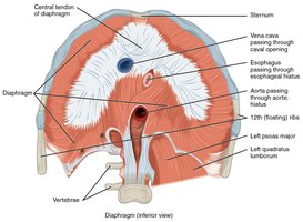
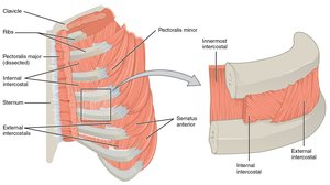
The diaphragm is the primary muscle of respiration, separating the thoracic and abdominal cavities. The intercostal muscles assist in breathing by moving the rib cage.
Diaphragm: Contracts to increase thoracic volume during inhalation.
External, Internal, and Innermost Intercostals: Elevate or depress the ribs during breathing.
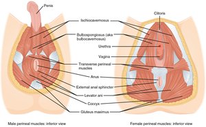
Muscles of the Pelvic Floor and Perineum
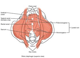
The pelvic floor muscles support pelvic organs, resist intra-abdominal pressure, and function as sphincters for the urethra, rectum, and vagina. The perineum muscles play roles in urination, ejaculation, and vaginal contraction.
Pelvic Floor: Levator ani, coccygeus.
Perineum: Bulbospongiosus, ischiocavernosus, transverse perineal muscles.
Muscles That Position the Pectoral Girdle and Move the Humerus
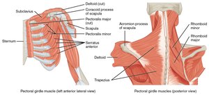
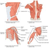
The muscles stabilizing the pectoral girdle provide a base for arm movement. Muscles that move the humerus originate from the trunk or scapula and insert into the humerus, allowing for a wide range of shoulder movements.
Pectoral Girdle: Trapezius, rhomboids, serratus anterior.
Humerus Movers: Deltoid, pectoralis major, latissimus dorsi, rotator cuff muscles.
Muscles That Move the Forearm, Wrist, and Hand
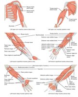
Muscles originating in the upper arm flex, extend, pronate, and supinate the forearm. Muscles in the forearm move the wrist, hand, and fingers, allowing for complex manipulations.
Forearm Movers: Biceps brachii, triceps brachii, brachialis, pronator teres, supinator.
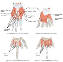
Wrist/Hand Movers: Flexor carpi radialis, extensor digitorum, intrinsic hand muscles.
Muscles of the Hip, Thigh, and Lower Leg
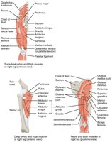
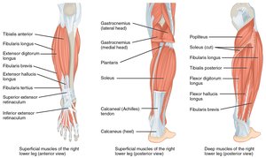
The hip and thigh muscles are large and powerful, responsible for moving the femur and lower leg. The lower leg muscles control movements of the ankle, foot, and toes.
Hip/Thigh Movers: Gluteus maximus, quadriceps femoris, hamstrings, adductors.
Lower Leg Movers: Tibialis anterior, gastrocnemius, soleus, fibularis muscles.
Intrinsic Muscles of the Foot
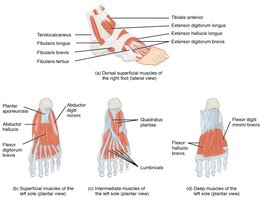
The intrinsic muscles of the foot originate and insert within the foot, providing fine motor control for toe movements and supporting the arches of the foot.
Dorsal Muscles: Extend the toes.
Plantar Muscles: Flex the toes and support the foot's arch.
Summary Table: Muscle Group Functions
The following table summarizes the main muscle groups, their primary actions, and examples:
Muscle Group | Main Action | Example Muscles |
|---|---|---|
Facial | Facial expression | Orbicularis oculi, orbicularis oris |
Mastication | Chewing | Masseter, temporalis |
Neck | Head movement | Sternocleidomastoid, trapezius |
Abdomen | Trunk movement, protection | Rectus abdominis, external oblique |
Pectoral Girdle | Shoulder stabilization | Trapezius, serratus anterior |
Upper Limb | Arm, forearm, hand movement | Biceps brachii, triceps brachii |
Hip/Thigh | Leg movement | Gluteus maximus, quadriceps |
Lower Leg/Foot | Foot and toe movement | Tibialis anterior, gastrocnemius |
Additional info: For a comprehensive understanding, students should also review the origin, insertion, action, and innervation of each muscle, as well as clinical correlations such as muscle injuries and disorders.
