 뒤로
The Muscular System: Structure, Organization, and Function
스터디 가이드 - 스마트 노트
 자료에 맞춘 맞춤형 노트, 핵심 정의, 예시, 맥락을 확장해 제공합니다.
The Muscular System
Introduction to the Muscular System
The muscular system is a complex network of nearly 700 muscles that enables movement, maintains posture, and supports bodily functions. The organization of muscle fibers into fascicles directly affects the power, range, and speed of muscle movement.
Muscle fascicles are bundles of muscle fibers (cells) that determine muscle function.
Muscle tension depends on the number of myofibrils and the cross-sectional area of the muscle.
Each square inch of muscle cross-section can generate approximately 50 pounds of tension.
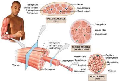
Muscle Fiber Structure
Muscle fibers contain myofibrils, which are the contractile units of the muscle.
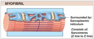
Myofibrils are composed of repeating units called sarcomeres, which are responsible for muscle contraction.
Myofibrils are surrounded by the sarcoplasmic reticulum, which regulates calcium ion concentration during contraction.
Fascicle Arrangement and Classification of Skeletal Muscle Fibers
Patterns of Fascicle Organization
The arrangement of fascicles within a muscle determines its function and mechanical properties. There are four primary patterns: parallel, convergent, pennate, and circular.
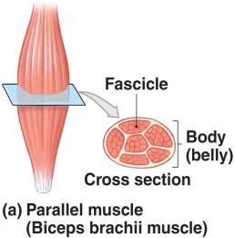
Parallel muscles: Fascicles run parallel to the long axis of the muscle. Example: Biceps brachii.
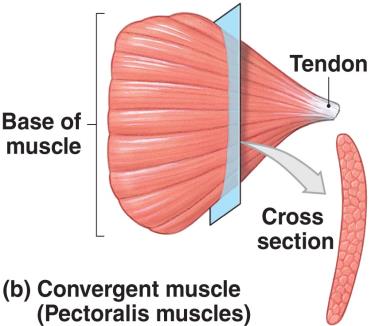
Convergent muscles: Fascicles converge from a broad area to a single attachment point (tendon, aponeurosis, or raphe). Example: Pectoralis major.
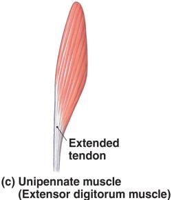
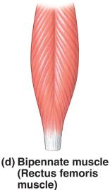
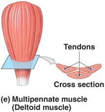
Pennate muscles: Fascicles are arranged at an angle to the tendon. These muscles generate more tension but less movement than parallel muscles. Types include:
Unipennate: Fascicles on one side of the tendon. Example: Extensor digitorum.
Bipennate: Fascicles on both sides of the tendon. Example: Rectus femoris.
Multipennate: Tendon branches within the muscle. Example: Deltoid.
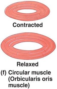
Circular muscles (sphincters): Fascicles are arranged concentrically around an opening, controlling its diameter. Example: Orbicularis oris.
Levers and Muscle Mechanics
Levers in Skeletal Motion
Skeletal muscles produce movement by acting on bones, which function as levers. Joints serve as fulcrums, and muscles provide the applied force to overcome resistance (load).
The function of a lever is to change the direction, distance, speed, or strength of the applied force.
There are three classes of levers, based on the arrangement of the fulcrum, applied force, and load:
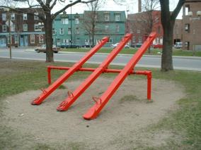
First-Class Lever
Fulcrum is positioned between the load and applied force.
Example: Seesaw, or the head moving on the neck.
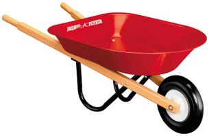
Second-Class Lever
Load is positioned between the fulcrum and applied force.
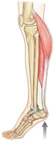
Example: Wheelbarrow, or standing on tiptoe.
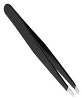
Third-Class Lever
Applied force is positioned between the fulcrum and load.
Most common lever type in the body; maximizes speed and distance.
Example: Flexing the forearm at the elbow.
Muscle Attachment and Actions
Origins and Insertions
Muscles attach to bones at two points: the origin (fixed, usually proximal) and the insertion (moving, usually distal).
Movement occurs when the insertion is pulled toward the origin.
Muscle Actions and Interactions
Muscle actions are described by the movement they produce (e.g., flexion, extension, adduction).
Muscles work in groups for efficiency; smaller muscles activate first, followed by larger ones.
Muscle roles:
Agonist (prime mover): Produces a specific movement.
Antagonist: Opposes the action of the agonist.
Synergist: Assists the agonist or stabilizes its origin.
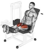
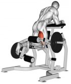
Agonists and antagonists often work in pairs (e.g., flexors and extensors).
Naming Skeletal Muscles
Muscle Naming Conventions
The names of skeletal muscles are based on several descriptive criteria:
Location: Indicates the region of the body (e.g., Genioglossus muscle from chin to tongue).
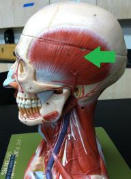
Origin and Insertion: The first part of the name is the origin, the second is the insertion.
Fascicle Organization: Describes the orientation of fascicles (e.g., Rectus = straight, Transverse = across, Oblique = angled).
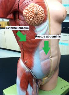
Relative Position: Indicates depth or location (e.g., Externus or superficialis = superficial; Internus or profundus = deep).
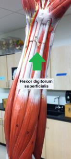
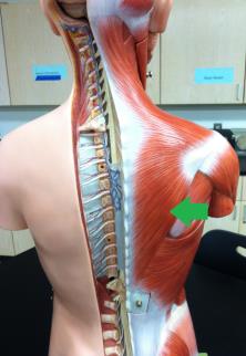
Structural Characteristics: Number of tendons (bi-, tri-), shape (trapezius, deltoid, rhomboid), and size (longus, brevis, magnus, maximus, minor, minimus, vastus).
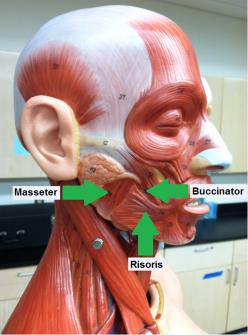
Action: Describes the movement produced (e.g., flexor, extensor, adductor) or habits (e.g., risor = laughter).
Divisions of the Muscular System
Axial and Appendicular Muscles
The muscular system is divided into two main groups:
Axial muscles: Position the head and spinal column, move the rib cage; comprise 60% of skeletal muscles.
Appendicular muscles: Support the pectoral and pelvic girdles and limbs; comprise 40% of skeletal muscles.
Summary Table: Fascicle Arrangement Types
Type | Arrangement | Example | Function |
|---|---|---|---|
Parallel | Fascicles parallel to long axis | Biceps brachii | High range of motion |
Convergent | Fascicles converge to a point | Pectoralis major | Versatile direction of pull |
Pennate | Fascicles at angle to tendon | Rectus femoris, deltoid | High tension, less movement |
Circular | Fascicles arranged in rings | Orbicularis oris | Control openings |
Summary Table: Muscle Roles
Role | Description | Example |
|---|---|---|
Agonist | Prime mover of action | Biceps brachii (elbow flexion) |
Antagonist | Opposes agonist | Triceps brachii (elbow extension) |
Synergist | Assists agonist | Brachialis (elbow flexion) |
Fixator | Stabilizes origin | Muscles stabilizing scapula |
Additional info: Expanded explanations and tables were added for clarity and completeness, based on standard anatomy and physiology textbook content.
