 뒤로
The Respiratory System: Structure and Function
스터디 가이드 - 스마트 노트
 자료에 맞춘 맞춤형 노트, 핵심 정의, 예시, 맥락을 확장해 제공합니다.
The Respiratory System
Overview and Major Functions
The respiratory system is essential for gas exchange, supplying oxygen (O2) to the body for cellular respiration and removing carbon dioxide (CO2), a waste product. It also plays roles in olfaction and speech. The processes of respiration involve both the respiratory and circulatory systems.
Pulmonary ventilation: Movement of air into and out of the lungs.
External respiration: Exchange of O2 and CO2 between lungs and blood.
Transport: Movement of O2 and CO2 in the blood.
Internal respiration: Exchange of O2 and CO2 between systemic blood vessels and tissues.
Functional Anatomy of the Respiratory System
Major Organs
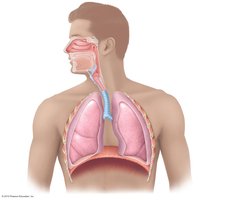
The respiratory system consists of several organs that facilitate the movement and exchange of gases:
Nose, nasal cavity, and paranasal sinuses
Pharynx
Larynx
Trachea
Bronchi and their branches
Lungs and alveoli
Zones of the Respiratory System
Respiratory zone: Site of gas exchange; includes respiratory bronchioles, alveolar ducts, and alveoli.
Conducting zone: Conduits to gas exchange sites; includes all other respiratory structures. Functions to cleanse, warm, and humidify air.
The Nose and Nasal Cavity
Structure and Functions
The nose provides an airway for respiration, moistens and warms incoming air, filters and cleans inspired air, serves as a resonating chamber for speech, and houses olfactory receptors.
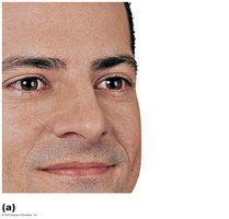
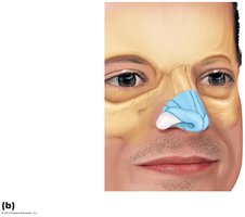
External nose: Includes the root, bridge, dorsum nasi, apex, philtrum, and nostrils (nares).
Nasal cavity: Located within and posterior to the external nose, divided by the nasal septum, and opens into the nasopharynx.
Nasal Cavity Regions and Mucosa
Nasal vestibule: Superior to nostrils; hairs filter coarse particles.
Olfactory mucosa: Contains olfactory epithelium for smell.
Respiratory mucosa: Pseudostratified ciliated columnar epithelium; mucous and serous secretions contain lysozyme and defensins.
Cilia: Move contaminated mucus posteriorly to the throat.
Capillaries and veins: Warm inspired air.
Sensory nerve endings: Trigger sneezing.
Functions of Nasal Mucosa and Conchae
During inhalation, conchae and nasal mucosa filter, heat, and moisten air.
During exhalation, these structures reclaim heat and moisture.
Paranasal Sinuses
Located in the frontal, sphenoid, ethmoid, and maxillary bones, paranasal sinuses lighten the skull, secrete mucus, and help warm and moisten air.
Homeostatic Imbalance: Rhinitis
Rhinitis is inflammation of the nasal mucosa. Because the nasal mucosa is continuous with the mucosa of the respiratory tract, infection can spread from the nose to the throat and chest, and to tear ducts and paranasal sinuses, causing blocked sinus passageways and sinus headaches.
Pharynx
Structure and Regions
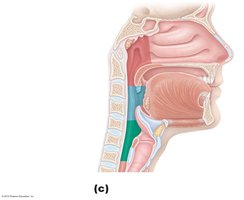
The pharynx is a muscular tube extending from the base of the skull to the level of the sixth cervical vertebra (C6). It connects the nasal cavity and mouth to the larynx and esophagus and is composed of skeletal muscle.
Nasopharynx
Oropharynx
Laryngopharynx
Larynx
Structure and Functions
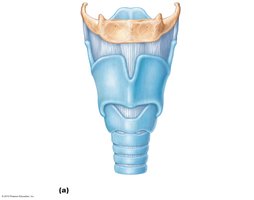
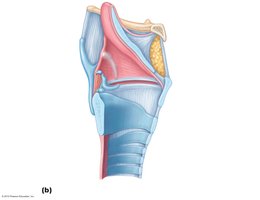
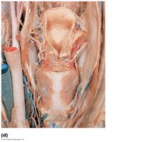
The larynx attaches to the hyoid bone, opens into the laryngopharynx, and is continuous with the trachea. It provides a patent airway, routes air and food into proper channels, produces voice, and houses the vocal folds.
Epiglottis: Prevents food from entering the airway.
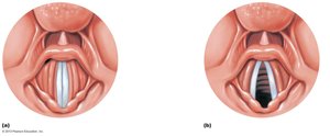
Vocal folds: Produce sound and may act as a sphincter to prevent air passage (e.g., during Valsalva's maneuver).
Trachea
Structure and Tissue Composition
The trachea, or windpipe, extends from the larynx into the mediastinum. Its wall is composed of three layers:
Mucosa: Ciliated pseudostratified epithelium with goblet cells.
Submucosa: Connective tissue with seromucous glands.
Adventitia: Outermost layer made of connective tissue, encasing C-shaped rings of hyaline cartilage.
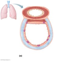
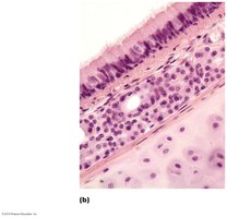
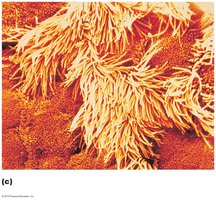
Trachealis Muscle and Carina
Trachealis muscle: Connects posterior parts of cartilage rings; contracts during coughing to expel mucus.
Carina: Spar of cartilage at the point where the trachea branches into two main bronchi.
Bronchi and Subdivisions
Bronchial Tree and Conducting Zone
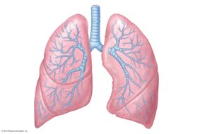
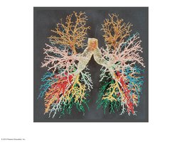
The bronchial tree undergoes 23 orders of branching, giving rise to conducting and respiratory zone structures. The trachea branches into right and left main (primary) bronchi, which enter the hilum of each lung. The right main bronchus is wider, shorter, and more vertical than the left.
Lobar (secondary) bronchi: Three on the right, two on the left; each supplies one lobe.
Segmental (tertiary) bronchi: Branches divide repeatedly, becoming smaller.
Bronchioles: Less than 1 mm in diameter.
Terminal bronchioles: Smallest, less than 0.5 mm in diameter.
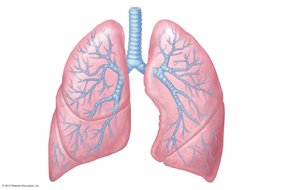
Structural Changes from Bronchi to Bronchioles
Cartilage rings become irregular plates; elastic fibers replace cartilage in bronchioles.
Epithelium changes from pseudostratified columnar to cuboidal; cilia and goblet cells become sparse.
Relative amount of smooth muscle increases, allowing constriction.
Respiratory Zone
Structure and Function
The respiratory zone begins as terminal bronchioles lead to respiratory bronchioles, alveolar ducts, and alveolar sacs. Alveolar sacs contain clusters of alveoli, which are the sites of gas exchange. Approximately 300 million alveoli make up most of the lung volume.
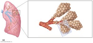
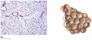
Alveoli and the Respiratory Membrane
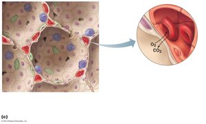
The respiratory membrane consists of alveolar and capillary walls and their fused basement membranes, approximately 0.5 μm thick. Gas exchange occurs across this membrane by simple diffusion.
Type I alveolar cells: Single layer of squamous epithelium.
Type II alveolar cells: Secrete surfactant and antimicrobial proteins.
Alveolar pores: Connect adjacent alveoli, equalizing air pressure throughout the lung.
Alveolar macrophages: Keep alveolar surfaces sterile.
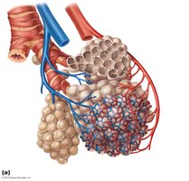
Lungs
Anatomical Features
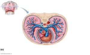
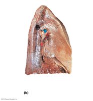
The lungs occupy all of the thoracic cavity except the mediastinum. The root is the site of vascular and bronchial attachment to the mediastinum. The costal surface includes anterior, lateral, and posterior surfaces. The lungs are composed primarily of alveoli.
Apex: Superior tip, deep to the clavicle.
Base: Inferior surface, rests on the diaphragm.
Hilum: Site for entry/exit of blood vessels, bronchi, lymphatic vessels, and nerves.
Left lung: Smaller than right, with a cardiac notch for the heart; separated into superior and inferior lobes by the oblique fissure.
Right lung: Superior, middle, and inferior lobes separated by oblique and horizontal fissures.
Bronchopulmonary segments: 10 in the right lung, 8–10 in the left; separated by connective tissue septa.
Lobules: Smallest subdivisions visible to the naked eye; served by bronchioles and their branches.
Blood Supply
Pulmonary and Bronchial Circulation
Pulmonary circulation: Low pressure, high volume; pulmonary arteries deliver deoxygenated blood to lungs for oxygenation; pulmonary veins carry oxygenated blood to the heart.
Lung capillary endothelium: Contains enzymes (e.g., angiotensin-converting enzyme) that act on substances in blood.
Bronchial arteries: Provide oxygenated blood to lung tissue (except alveoli); arise from the aorta and enter lungs at the hilum; part of systemic circulation (high pressure, low volume).
Pleurae
Structure and Function
The pleurae are thin, double-layered serous membranes that divide the thoracic cavity into two pleural compartments and the mediastinum.
Parietal pleura: Lines the thoracic wall, superior face of the diaphragm, around the heart, and between the lungs.
Visceral pleura: Covers the external lung surface.
Pleural fluid: Fills the slitlike pleural cavity, providing lubrication and surface tension to assist in expansion and recoil of the lungs.
