 뒤로
The Skeletal System: Structure, Function, and Classification
스터디 가이드 - 스마트 노트
 자료에 맞춘 맞춤형 노트, 핵심 정의, 예시, 맥락을 확장해 제공합니다.
The Skeletal System
Overview of the Skeletal System
The skeletal system is a complex framework of bones that provides structural support, protection, and movement for the human body. It consists of approximately 206 bones, which are classified into two main divisions: the axial skeleton and the appendicular skeleton.
Axial skeleton: Includes the skull, vertebral column, and thoracic cage. It forms the central axis of the body and protects vital organs.
Appendicular skeleton: Comprises the girdles and limbs, facilitating movement and interaction with the environment.
Classification of Bones
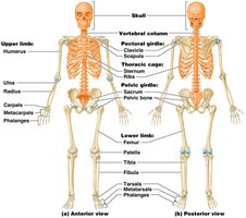
Bones are classified based on their location and function within the skeletal system. The axial skeleton includes the skull, vertebral column, and thoracic cage, while the appendicular skeleton consists of the pectoral and pelvic girdles, as well as the upper and lower limbs.
Skull: Protects the brain and forms the structure of the face.
Vertebral column: Protects the spinal cord and supports the body.
Thoracic cage: Protects the heart and lungs.
Pectoral girdle: Anchors the upper limbs to the trunk.
Pelvic girdle: Anchors the lower limbs to the trunk.
Bone Markings
Types of Bone Markings
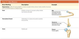
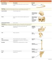
Bone markings are structural features on bones that serve as sites for muscle attachment, passage of nerves and blood vessels, and articulation with other bones. They are classified as depressions, openings, and projections.
Depressions: Include facets, fossae, and grooves, which allow for articulation or passage of structures.
Openings: Include foramina, canals, and fissures, which permit the passage of nerves and blood vessels.
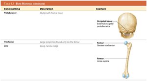
Projections: Include processes, trochanters, and lines, which serve as attachment points for muscles and ligaments.
Bone Marking | Description | Example |
|---|---|---|
Facet | Shallow convex or concave surface | Articular facet for rib |
Fossa | Indentation in a bone | Distal portion of humerus |
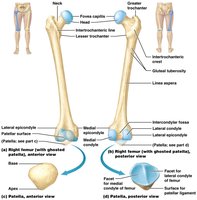
Fovea | Shallow pit | Fovea capitis of femur |
Foramen | Hole in a bone | Foramen magnum of occipital bone |
Process | Prominent bony projection | Mastoid process of temporal bone |
Trochanter | Large projection found only on femur | Greater trochanter of femur |
Line | Long, narrow ridge | Linea aspera of femur |
The Skull
Cranial and Facial Bones
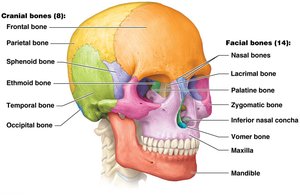
The skull is composed of 22 bones, divided into cranial and facial groups. Cranial bones protect the brain, while facial bones form the structure of the face.
Cranial bones (8): Frontal, parietal, sphenoid, ethmoid, temporal, and occipital bones.
Facial bones (14): Nasal, lacrimal, palatine, zygomatic, inferior nasal concha, vomer, maxilla, and mandible.
Sutures of the Skull
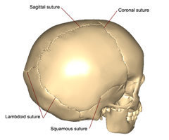
Most bones of the skull are fused at immovable joints called sutures. Four major sutures unite specific cranial bones.
Coronal suture: Unites the frontal and parietal bones.
Sagittal suture: Unites the two parietal bones.
Lambdoid suture: Unites the parietal and occipital bones.
Squamous suture: Unites the parietal and temporal bones.
Paranasal Sinuses
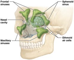
Paranasal sinuses are air-filled cavities within certain skull bones. They are lined with mucous membranes that filter, warm, and humidify inspired air, reduce skull weight, and enhance voice resonance.
Sinuses are found in: Frontal, sphenoid, ethmoid, and maxillary bones.
The Fetal Skull and Fontanels
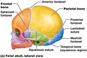
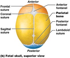
In the fetal skull, bones are not fused, and membranous areas called fontanels are present. Fontanels allow flexibility during birth and accommodate brain growth.
Fontanels: Anterior, posterior, sphenoid, and mastoid fontanels.
Importance: Permit passage through the birth canal and expansion with brain growth.
Hyoid Bone
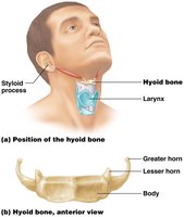
The hyoid bone is a unique, C-shaped bone in the neck that does not articulate with any other bone. It is suspended by ligaments and provides attachment for muscles involved in swallowing and speech.
Gender Differences in the Skull
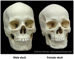
Male and female skulls exhibit distinct anatomical differences, which can be used for identification.
Male skull: Sloped forehead, prominent supraorbital ridge, mandibular angle closer to 90°, larger mastoid process.
Female skull: Straight forehead, less prominent supraorbital ridge, mandibular angle greater than 90°, smaller mastoid process.
The Vertebral Column
Classification of Vertebrae
The vertebral column consists of 33 vertebrae, classified by region and function.
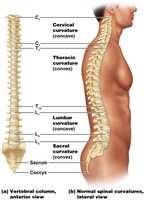
Cervical (7): Located in the neck; C1 (Atlas) and C2 (Axis) are specialized for head movement.
Thoracic (12): Articulate with ribs.
Lumbar (5): Located in the lower back.
Sacral (5 fused): Form the sacrum, articulating with pelvic bones.
Coccygeal (3–5 fused): Form the coccyx, the most inferior part.
Spinal Curvatures
The vertebral column exhibits four curvatures, which are classified as primary (present at birth) and secondary (develop after birth).
Primary curvatures: Thoracic and sacral.
Secondary curvatures: Cervical and lumbar.
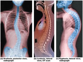
Abnormal Spinal Curvatures
Abnormal curvatures of the spine include:
Scoliosis: Abnormal lateral curvature.
Lordosis: Exaggerated cervical and lumbar curvatures (swayback).
Kyphosis: Exaggeration of thoracic curvature (hunchback).
Intervertebral Discs
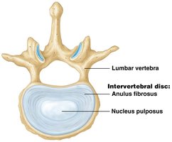
Intervertebral discs are fibrocartilage pads located between vertebrae. They absorb shock and bind the vertebral column together.
Anulus fibrosus: Tough outer layer.
Nucleus pulposus: Gel-like inner core.
The Thoracic Cage
Structure and Function
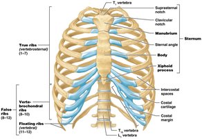
The thoracic cage consists of the sternum, 12 pairs of ribs, and thoracic vertebrae. It protects the thoracic cavity and vital organs.
True ribs (1–7): Attach directly to the sternum via costal cartilages.
False ribs (8–10): Attach indirectly to the sternum.
Floating ribs (11–12): Do not attach to the sternum.
Intercostal spaces: Gaps between ribs.
The Pectoral Girdle and Upper Limb
Pectoral Girdle
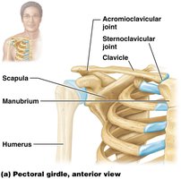
The pectoral girdle consists of the clavicle and scapula, anchoring the upper limb to the trunk. The glenoid cavity is a key feature for articulation with the humerus.
Upper Limb Bones
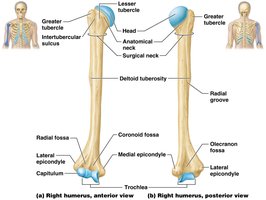
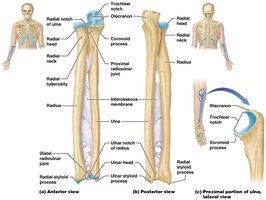
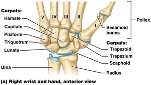
The upper limb includes the humerus, radius, ulna, carpals, metacarpals, and phalanges.
Humerus: The bone of the upper arm.
Radius and ulna: Bones of the forearm.
Carpals: Wrist bones.
Metacarpals: Bones of the hand.
Phalanges: Finger bones.
The Pelvic Girdle and Lower Limb
Pelvic Girdle and Pelvis
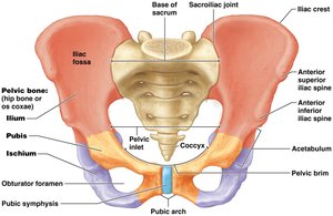
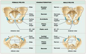
The pelvic girdle is composed of two coxal bones. The pelvis includes the pelvic girdle plus the sacrum. The acetabulum is the socket for the femur.
Gender Differences in the Pelvis
There are notable differences between male and female pelvises, primarily related to childbirth.
Female pelvis: Wider, shallower, flared iliac crests, wider sacrum and coccyx, more moveable coccyx, wider and oval-shaped inlet, wider outlet, greater pubic arch angle.
Male pelvis: Narrower, deeper, heart-shaped inlet, narrower outlet, smaller pubic arch angle.
Characteristic | Female Pelvis | Male Pelvis |
|---|---|---|
Pelvic inlet | Wide, oval | Narrow, heart-shaped |
Pelvic outlet | Wide | Narrow |
Pubic arch | Greater angle | Smaller angle |
Sacrum | Wide, short | Narrow, long |
Coccyx | More moveable | Less moveable |
Lower Limb Bones
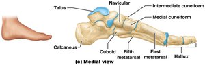
The lower limb includes the femur, patella, tibia, fibula, tarsals, metatarsals, and phalanges.
Femur: Largest and strongest bone in the body.
Patella: Kneecap.
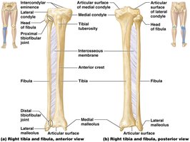
Tibia and fibula: Bones of the leg.
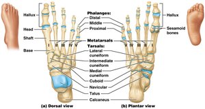
Tarsals: Ankle bones.
Metatarsals: Bones of the foot.
Phalanges: Toe bones.
Summary Table: Major Bones of the Human Skeleton
Region | Main Bones |
|---|---|
Skull | Cranial (8), Facial (14) |
Vertebral Column | Cervical (7), Thoracic (12), Lumbar (5), Sacrum (5 fused), Coccyx (3–5 fused) |
Thoracic Cage | Sternum, Ribs (12 pairs) |
Pectoral Girdle | Clavicle, Scapula |
Upper Limb | Humerus, Radius, Ulna, Carpals, Metacarpals, Phalanges |
Pelvic Girdle | Coxal bones |
Lower Limb | Femur, Patella, Tibia, Fibula, Tarsals, Metatarsals, Phalanges |
Additional info: The notes above include expanded academic context and logical grouping of fragmented topics to ensure completeness and clarity for exam preparation.
