 뒤로
The Skeletal System: Structure, Function, and Classification
스터디 가이드 - 스마트 노트
 자료에 맞춘 맞춤형 노트, 핵심 정의, 예시, 맥락을 확장해 제공합니다.
The Skeletal System
Overview and Functions
The skeletal system is a complex framework of bones and connective tissues that provides structural support, protection, movement, blood cell production, and storage of minerals and fats.
Support: Forms the structural framework for the body, maintaining posture.
Protection: Shields vital organs (e.g., skull protects the brain, ribcage protects heart and lungs).
Movement: Bones act as levers in conjunction with muscles, tendons, and joints.
Blood Cell Production: Bone marrow produces red and white blood cells and platelets.
Mineral and Fat Storage: Stores calcium, phosphorus, and fat in yellow bone marrow.
Types of Bone Tissue
Bone is a hard, dense connective tissue (osseous tissue) with a calcified matrix, while cartilage is softer, flexible, and avascular.
Bone: Contains osteocytes, osteoblasts, and osteoclasts; classified as compact or spongy bone.
Cartilage: Contains chondrocytes; classified as hyaline, elastic, or fibrocartilage.
Divisions of the Skeletal System
The human skeleton is divided into two main regions:
Axial Skeleton: Skull, spinal column, thoracic cage.
Appendicular Skeleton: Pectoral girdle, upper limbs, pelvic girdle, lower limbs.
Total Bones: 206 in the adult human skeleton.
Classification of Bones
Types and Examples
Long Bones: Longer than wide; femur, humerus, tibia, fibula, radius, ulna, clavicle, phalanges.
Short Bones: Cube-shaped; carpal (wrist) and tarsal (ankle) bones.
Flat Bones: Thin, broad surfaces; cranial bones, sternum, ribs, scapula.
Irregular Bones: Complex shapes; vertebrae, sacrum, coccyx, hyoid, certain facial bones.
Surface Markings
Form joints with other bones.
Provide anchor points for muscles and connective tissue.
Allow passage for blood vessels and nerves.
Structure of a Long Bone
Main Regions and Internal Structures
Long bones have specialized regions and internal structures for strength and mobility:
Diaphysis: Central shaft, made of compact bone.
Epiphysis: Rounded ends, form joints.
Metaphysis: Transition zone, contains growth plate in children.
Articular Cartilage: Covers epiphyses, reduces friction.
Periosteum: Outer membrane, supports growth and repair.
Compact Bone: Dense, hard outer layer.
Spongy Bone: Porous inner layer, mainly in epiphyses.
Medullary Cavity: Hollow central space in diaphysis.
Endosteum: Lines medullary cavity and inner surfaces.
Bone Marrow: Red marrow produces blood cells; yellow marrow stores fat.
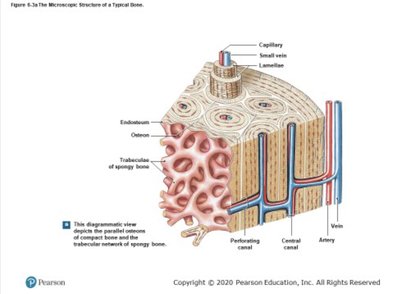
Microscopic Structure of Bone
Key Components
Lamellae: Concentric rings of bone matrix.
Osteons: Structural units of compact bone.
Trabeculae: Network in spongy bone.
Central Canal: Contains blood vessels and nerves.
Perforating Canal: Horizontal channels for vessels.
Endosteum: Membrane lining internal surfaces.
Capillary, Artery, Vein: Vascular supply for bone tissue.
Vertebral Column and Spine
Regions and Functions
Cervical Spine: 7 vertebrae (C1–C7); C1 (Atlas) and C2 (Axis) allow head movement.
Thoracic Spine: 12 vertebrae (T1–T12).
Lumbar Spine: 5 vertebrae (L1–L5).
Sacrum: 5 fused vertebrae.
Coccyx: 4 fused vertebrae.
Functions: Structural support, spinal cord protection, movement, flexibility, shock absorption.
Structural Parts of Vertebrae
Vertebral Body: Weight-bearing portion.
Vertebral Arch: Encloses spinal cord; made of pedicles and laminae.
Vertebral Foramen: Central opening for spinal cord.
Rib Classification and Articulation
Types of Ribs
True Ribs (1–7): Directly attach to sternum via costal cartilage.
False Ribs (8–10): Indirect attachment via cartilage of rib above.
Floating Ribs (11–12): No anterior attachment.
Ribs articulate posteriorly with the vertebral column and anteriorly with the sternum or cartilage.
Spinal Curves
Developmental Curves
Primary Curves: Thoracic and sacral curves, present at birth (concave anteriorly).
Secondary Curves: Cervical and lumbar curves, develop after birth (convex anteriorly).
Bone Growth and Remodeling
Appositional Growth
Appositional growth increases bone thickness or diameter by adding new layers to the outer surface.
Bone vs. Cartilage
Feature | Bone | Cartilage |
|---|---|---|
Rigidity | Hard, calcified | Soft, flexible |
Blood Supply | Rich, heals quickly | Avascular, slow healing |
Cell Types | Osteocytes, osteoblasts, osteoclasts | Chondrocytes |
Types | Compact, spongy | Hyaline, elastic, fibrocartilage |
Growth | Bidirectional | Unidirectional |
Function | Support, protection, movement, mineral storage | Shock absorption, flexibility |
Bone Cells and Their Roles
Osteoblasts, Osteocytes, Osteoclasts
Osteoblasts: Build new bone matrix; derived from mesenchymal stem cells.
Osteocytes: Maintain bone tissue, sense stress; former osteoblasts embedded in matrix.
Osteoclasts: Break down bone tissue; derived from hematopoietic stem cells.
Joints and Body Movements
Synovial Joints
Synovial joints are freely movable, characterized by a fluid-filled cavity and a fibrous capsule.
Articular Cartilage: Reduces friction, absorbs shock.
Articular Capsule: Encloses joint cavity; outer fibrous layer and inner synovial membrane.
Synovial Fluid: Lubricates, absorbs shock, nourishes cartilage.
Ligaments: Stabilize and align bones.
Types of Synovial Joints
Type | Location | Movement |
|---|---|---|
Ball-and-Socket | Shoulder, Hip | All directions, rotation |
Hinge | Elbow, Knee, Fingers/Toes | Flexion, extension |
Pivot | Neck, Forearm | Rotation |
Condyloid | Wrist, Knuckles | Side-to-side, up-and-down |
Saddle | Thumb base | Flexion, extension, abduction, adduction |
Plane (Gliding) | Wrist, Ankle, Spine | Sliding, gliding |
Body Movements
Flexion/Extension: Decrease/increase angle between body parts.
Abduction/Adduction: Move away/toward midline.
Medial/Lateral Rotation: Rotate toward/away from midline.
Elevation/Depression: Move upward/downward.
Protraction/Retraction: Move forward/backward.
Specific Bones of the Skeleton
Skull Bones
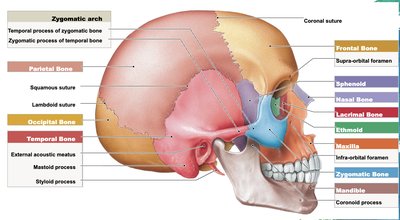
The adult skull is divided into cranial and facial bones:
Cranial Bones (8): Frontal, parietal (2), temporal (2), occipital, sphenoid, ethmoid.
Facial Bones (14): Mandible, maxillae (2), zygomatic (2), nasal (2), lacrimal (2), palatine (2), vomer, inferior nasal conchae (2).
Pelvic Girdle
Hip Bones: Ilium, ischium, pubis (fused in adults).
Sacrum: 5 fused vertebrae.
Coccyx: 4 fused vertebrae.
Pectoral Girdle
Clavicle: Collarbone (2).
Scapula: Shoulder blade (2).
Sternum and Thorax
Sternum: Manubrium, body, xiphoid process.
Ribs: 24 total (12 pairs: 7 true, 3 false, 2 floating).
