 뒤로
뒤로The Skeleton & Joints: Structure, Function, and Clinical Correlations
스터디 가이드 - 스마트 노트
 자료에 맞춘 맞춤형 노트, 핵심 정의, 예시, 맥락을 확장해 제공합니다.
Organization of the Skeletal System
Axial and Appendicular Skeleton
The human skeleton is divided into two main parts: the axial skeleton and the appendicular skeleton. The axial skeleton forms the long axis of the body and includes the skull, vertebral column, and rib cage. The appendicular skeleton consists of the bones of the upper and lower limbs and the girdles that attach them to the axial skeleton.
Axial skeleton: Skull, vertebral column, rib cage
Appendicular skeleton: Limbs and girdles (shoulder and pelvic)
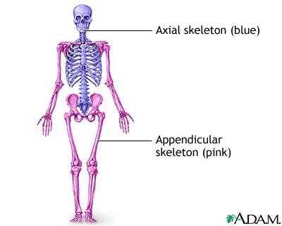
The Axial Skeleton
Vertebral (Spinal) Column
The vertebral column protects the spinal cord, supports the head, and transmits the weight of the trunk to the lower extremities. It also serves as an attachment point for the ribs, pelvic girdle, and muscles of the back. The column is a flexible, curved structure composed of 26 irregular bones called vertebrae.
Functions: Protection, support, weight transmission, muscle attachment
Structure: 26 vertebrae divided into regions
Regions of the Vertebral Column
Cervical: 7 vertebrae (neck)
Thoracic: 12 vertebrae (upper back)
Lumbar: 5 vertebrae (lower back)
Sacrum: 1 bone (fusion of several vertebrae, articulates with hip)
Coccyx: 1 bone (fusion of several small vertebrae, forms the terminus)
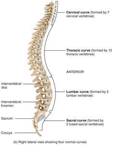
Vertebral Column Curves
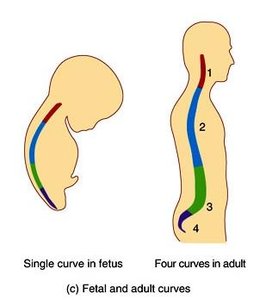
The vertebral column has four main curves that increase its strength, aid in shock absorption, help maintain upright balance, and protect vertebrae from fracture.
Primary curves: Thoracic and sacral (present at birth)
Secondary curves: Cervical and lumbar (develop after birth, complete by age 10)
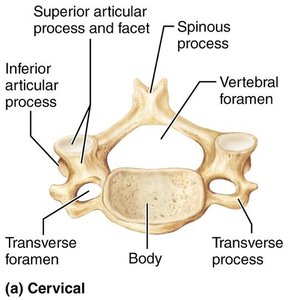
Characteristics of Cervical, Thoracic, and Lumbar Vertebrae
Each region of the vertebral column has vertebrae with distinct characteristics:
Cervical: Small, oval body; bifid spinous process; large, triangular vertebral foramen; greatest range of movement
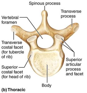
Thoracic: Larger, heart-shaped body; long, sharp spinous process; circular vertebral foramen; rotation and limited flexion/extension
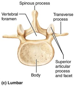
Lumbar: Massive, kidney-shaped body; short, blunt spinous process; triangular vertebral foramen; flexion/extension, limited rotation
Abnormal Vertebral Column Curves
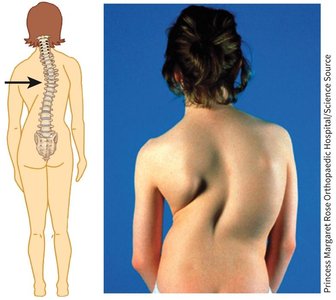
Scoliosis: Lateral bending of the vertebral column; causes include congenital malformation, muscle paralysis, poor posture, or leg length discrepancy.
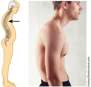
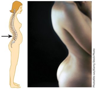
Kyphosis: Exaggerated thoracic curve; causes include osteoporosis, poor posture, or disc degeneration.
Lordosis: Exaggerated lumbar curve; causes include increased abdominal weight, poor posture, or rickets.
Intervertebral Discs
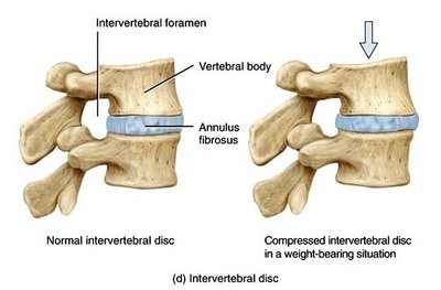
Intervertebral discs are cushion-like pads between vertebrae that act as shock absorbers. Each disc has two main parts:
Nucleus pulposus: Inner gelatinous core, provides elasticity and compressibility
Anulus fibrosus: Outer collar of collagen and fibrocartilage, limits expansion of nucleus pulposus
Herniated Discs
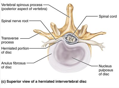
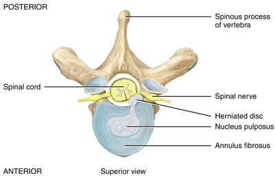
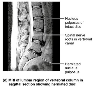
Increased stress can cause the nucleus pulposus to rupture through the anulus fibrosus, most often in the lumbar region. This can compress spinal nerves, causing pain, muscle weakness, or atrophy.
Thoracic Cage
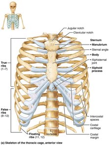
The thoracic cage is composed of the thoracic vertebrae, ribs, sternum, and costal cartilages. It protects vital organs, supports the shoulder girdles and upper limbs, and provides attachment sites for muscles.
True ribs (1–7): Attach directly to sternum
False ribs (8–10): Attach indirectly via costal cartilage
Floating ribs (11–12): No anterior attachment
The Appendicular Skeleton
Pectoral Girdle and Upper Limb
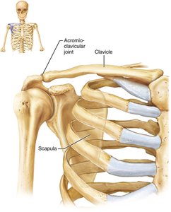
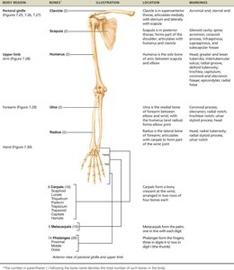
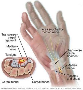
The pectoral girdle consists of the clavicles and scapulae, attaching the upper limbs to the axial skeleton. The upper limb includes the humerus (arm), radius and ulna (forearm), and the bones of the hand (carpals, metacarpals, phalanges).
Pelvic Girdle and Lower Limb
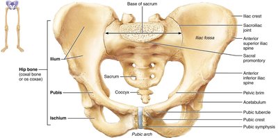
The pelvic girdle is formed by two hip bones and the sacrum, attaching the lower limbs to the axial skeleton. The lower limb includes the femur (thigh), tibia and fibula (leg), and the bones of the foot (tarsals, metatarsals, phalanges).
Joints (Articulations)
Definition and Classification
A joint (articulation or arthrosis) is the point of contact between two bones, bone and cartilage, or bone and teeth. Joints are classified by structure and function.
Structural classification: Based on the type of connective tissue and presence/absence of a synovial cavity
Functional classification: Based on the degree of movement permitted
Structural Classification of Joints
Fibrous joints: Bones joined by dense irregular connective tissue; no synovial cavity; little/no movement
Cartilaginous joints: Bones joined by cartilage; no synovial cavity; little/no movement
Synovial joints: Bones separated by a synovial cavity; freely movable
Functional Classification of Joints
Synarthroses: Immovable joints
Amphiarthroses: Slightly movable joints
Diarthroses: Freely movable joints
Fibrous Joints
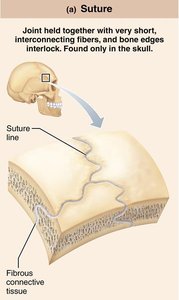
Fibrous joints are held together by dense irregular connective tissue and lack a synovial cavity. They allow little to no movement.
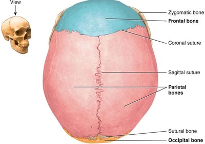
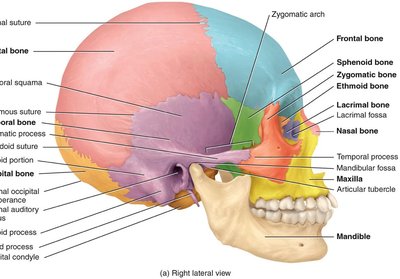
Sutures: Found only in the skull; immovable in adults
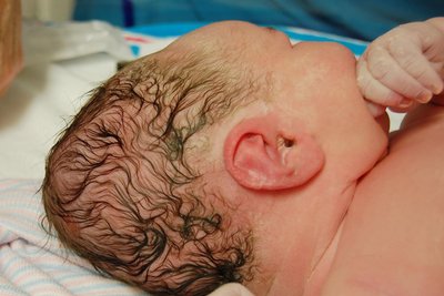
Fontanels: Amphiarthrotic in infants, allowing for growth of the skull
Syndesmoses: Bones connected by ligaments; allow slight movement (e.g., distal tibiofibular joint)
Interosseous membranes: Sheets of connective tissue joining long bones (e.g., between radius and ulna)
Cartilaginous Joints
Cartilaginous joints are tightly connected by hyaline cartilage or fibrocartilage and lack a synovial cavity. They allow little to no movement.
Synchondroses: Bones joined by hyaline cartilage (e.g., epiphyseal plates in children)
Symphyses: Bones joined by fibrocartilage (e.g., pubic symphysis, intervertebral discs)
Synovial Joints
Synovial joints are diarthroses (freely movable) and have a complex structure including an articular capsule, synovial cavity, synovial fluid, articular cartilage, ligaments, nerves, and blood vessels.
Articular capsule: Encloses the joint cavity, permits movement, and prevents dislocation
Synovial fluid: Lubricates, absorbs shock, supplies nutrients, and removes wastes
Articular cartilage: Hyaline cartilage covering bone surfaces, reduces friction and absorbs shock
Accessory structures: Ligaments, articular discs (menisci), labra, bursae, and tendon sheaths
Types of Synovial Joints
Plane (gliding): Biaxial movement
Hinge: Uniaxial movement (e.g., elbow)
Pivot: Uniaxial rotation (e.g., radioulnar joint)
Condyloid: Biaxial movement
Saddle: Biaxial movement
Ball and socket: Triaxial movement (e.g., shoulder, hip)
Movements at Synovial Joints
Flexion, extension, hyperextension
Abduction, adduction
Rotation (internal, external)
Special movements: Supination, pronation
Knee Joint: Structure and Clinical Correlations
The knee joint is a complex synovial joint involving the femur, tibia, and patella. It contains several ligaments (ACL, PCL, LCL, MCL), menisci, and bursae. Common injuries include ACL tears, meniscus tears, and patellofemoral syndrome.
ACL: Prevents anterior displacement of tibia; taut during extension
PCL: Prevents posterior displacement of tibia; taut during flexion
Menisci: Fibrocartilage discs that improve fit and distribute synovial fluid
Bursitis: Inflammation of a bursa due to repetitive use or trauma
Aging and Joints
Decreased synovial fluid production
Thinner articular cartilage
Shortened, less elastic ligaments
Increased risk of joint degeneration and fracture
Arthroplasty
Arthroplasty is the surgical replacement of a joint, most commonly the hip, knee, or shoulder. The goals are to relieve pain and increase range of motion. Complications include infection, blood clots, nerve injury, and prosthesis degeneration.
Summary Table: Structural and Functional Classification of Joints
Structural Type | Examples | Movement (Functional) |
|---|---|---|
Fibrous (Suture) | Skull sutures | Synarthrosis (immovable) |
Fibrous (Syndesmosis) | Distal tibiofibular joint | Amphiarthrosis (slightly movable) |
Cartilaginous (Synchondrosis) | Epiphyseal plate | Synarthrosis/Amphiarthrosis |
Cartilaginous (Symphysis) | Pubic symphysis, intervertebral discs | Amphiarthrosis |
Synovial (Hinge) | Elbow, knee | Diarthrosis (freely movable) |
Synovial (Ball and Socket) | Shoulder, hip | Diarthrosis |
