 뒤로
The Skeleton: Thoracic Cage and Pectoral Girdle
스터디 가이드 - 스마트 노트
 자료에 맞춘 맞춤형 노트, 핵심 정의, 예시, 맥락을 확장해 제공합니다.
The Skeleton: Thoracic Cage and Pectoral Girdle
Thoracic Cage
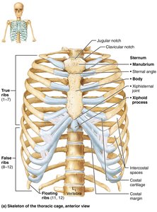
The thoracic cage is a vital bony structure of the chest, providing protection, support, and muscle attachment. It is composed of the thoracic vertebrae, ribs, sternum, and costal cartilages.
Components: Posteriorly: thoracic vertebrae; laterally: ribs; anteriorly: sternum and costal cartilages.
Functions: Protects vital organs (heart, lungs), supports shoulder girdles and upper limbs, and provides muscle attachment sites for the neck, back, chest, and shoulders.
Bones of the Thoracic Cage
Sternum: Also called the breastbone, consists of three fused bones:
Manubrium: Superior portion, articulates with clavicular notches and ribs 1 and 2.
Body: Midportion, articulates with costal cartilages of ribs 2–7.
Xiphoid process: Inferior end, site of muscle attachment; not ossified until ~ age 40.
Anatomical Landmarks:
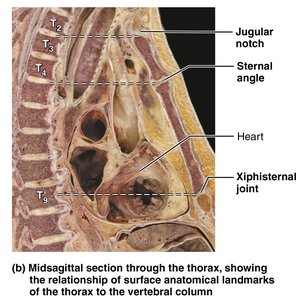
Jugular notch: Central indentation in superior border of manubrium.
Sternal angle: Horizontal ridge across front of sternum.
Xiphisternal joint: Point where sternal body and xiphoid process fuse.
Clinical Significance
Xiphoid process: Projects posteriorly in some people; chest trauma can push it into underlying liver or heart, causing hemorrhaging.
CPR: Hands must be positioned on the midsternum to avoid breaking off the xiphoid process and damaging underlying organs.
Ribs
The ribs form the sides of the thoracic cage and are classified based on their attachment to the sternum.
12 pairs of ribs: All attach posteriorly to bodies and transverse processes of thoracic vertebrae.
True (vertebrosternal) ribs (1–7): Attach directly to sternum by individual costal cartilages.
False (vertebrochondral) ribs (8–10): Attach indirectly to sternum by joining costal cartilage of rib above.
Floating (vertebral) ribs (11–12): No attachment to sternum.
Main Parts of a Rib
Shaft: Flat bone making up most of the rib.
Head: Posterior end, articulates with facets (demifacets) on bodies of two adjacent vertebrae.
Neck: Constricted portion beyond head.
Tubercle: Knoblike structure lateral to neck, articulates with transverse costal facet of same-numbered thoracic vertebra.


The Appendicular Skeleton: Pectoral Girdle
The appendicular skeleton consists of the bones of the limbs and their girdles. The pectoral girdle attaches the upper limbs to the body trunk and provides mobility and muscle attachment sites.
Pectoral girdle: Consists of clavicles (anteriorly) and scapulae (posteriorly).
Function: Attaches upper limbs to axial skeleton, provides attachment sites for muscles, and offers great mobility due to shallow shoulder joint socket and unattached scapulae.
Clavicle
Also called collarbone: S-shaped sternal end articulates with sternum medially; flattened acromial end articulates laterally with scapula.
Function: Anchors muscles and acts as braces to hold the scapulae and arms out laterally.
Scapula
Also called shoulder blade: Thin, triangular, irregular bone on dorsal surface of rib cage, between ribs 2 and 7.
Borders: Superior (shortest, sharpest), medial (vertebral, parallel to spine), lateral (axillary, near armpit, ends in glenoid cavity fossa).
Angles: Superior (between superior and medial), lateral (between superior and lateral), inferior (between medial and lateral).
Features: Spine (prominent ridge posteriorly), acromion (lateral projection, forms acromioclavicular joint), coracoid process (anterior projection, anchors biceps), suprascapular notch (opening for nerves), several large fossae (subscapular, supraspinous, infraspinous).
The Upper Limb
The upper limb consists of the arm, forearm, and hand, with a total of 30 bones forming its skeletal framework.
Arm: Humerus is the only bone, largest and longest in the upper limb. Articulates superiorly with glenoid cavity of scapula and inferiorly with radius and ulna.
Forearm: Two parallel bones: ulna (medial) and radius (lateral). Proximal ends articulate with humerus and each other; distal ends articulate with each other at the radioulnar joint. Interosseous membrane connects them along their length.
Hand: Includes carpus (wrist, 8 bones in two rows), metacarpus (palm, 5 bones), and phalanges (fingers, 14 bones).
Clinical Significance
Colles’ fracture: Break in distal end of radius, common when falling and attempting to break the fall with outstretched hands.
Carpal tunnel syndrome: Median nerve and tendons travel through carpal tunnel formed by ligaments; overuse and inflammation can compress the median nerve, causing tingling and numbness.
Table: Bones of the Appendicular Skeleton (Pectoral Girdle and Upper Limb)
Region | Bones | Number | Key Features |
|---|---|---|---|
Pectoral Girdle | Clavicle, Scapula | 2 each | Attachment to axial skeleton, muscle attachment |
Arm | Humerus | 1 | Largest bone, articulates with scapula, radius, ulna |
Forearm | Ulna, Radius | 2 each | Form elbow joint, radioulnar joint, interosseous membrane |
Hand | Carpals, Metacarpals, Phalanges | 8, 5, 14 | Wrist, palm, fingers |
Additional info: The notes expand on brief points from the slides, providing definitions, anatomical context, and clinical relevance for each structure. Images included are directly relevant to the anatomical structures discussed and reinforce the educational content.
