뒤로
The Spinal Cord, Spinal Nerves, and Spinal Reflexes: Structured Study Notes
스터디 가이드 - 스마트 노트
 자료에 맞춘 맞춤형 노트, 핵심 정의, 예시, 맥락을 확장해 제공합니다.
The Nervous System: Structural and Organizational Characteristics
Central and Peripheral Nervous System
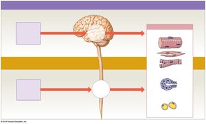
The nervous system is divided into the central nervous system (CNS) and the peripheral nervous system (PNS). The CNS consists of the brain and spinal cord, serving as the main processing centers. The PNS includes cranial and spinal nerves, which connect the CNS to the rest of the body and are responsible for sensory input and motor response pathways.
Central Nervous System (CNS): Brain and spinal cord; processes and integrates information.
Peripheral Nervous System (PNS): Cranial and spinal nerves; transmits sensory and motor signals.
Reflexes: Quick, automatic responses to stimuli, often processed at the spinal cord level without brain involvement.
Example: Dropping a hot pan before consciously feeling pain.
The Spinal Cord: Structure and Functions
Gross Anatomy and Regions
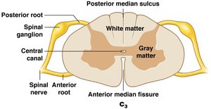
The spinal cord is a cylindrical structure housed within the vertebral column and protected by three meningeal layers. It carries sensory and motor information between the brain and the body, and gives rise to 31 pairs of spinal nerves.
Length: About 45 cm (18 in.) long, 14 mm (1/2 in.) wide.
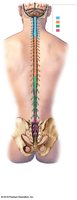
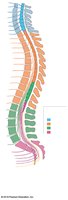
Regions: Cervical, thoracic, lumbar, sacral.
Segments: 31, each giving rise to a pair of spinal nerves.
Grooves: Posterior median sulcus (back), anterior median fissure (front).
Central canal: Contains cerebrospinal fluid (CSF).
Enlargements and Distal End
Cervical enlargement: Supplies shoulder and upper limb.
Lumbosacral enlargement: Supplies pelvis and lower limb.nm
Conus medullaris: Tapered end of the spinal cord below lumbar enlargement.
Cauda equina: Bundle of nerve roots below the conus medullaris.
Filum terminale: Fibrous tissue anchoring the cord to the coccyx.
Spinal Roots, Ganglia, and Nerves
Anterior (ventral) root: Motor neuron axons.
Posterior (dorsal) root: Sensory neuron axons.
Spinal ganglia: Contain sensory neuron cell bodies.
Spinal nerves: Formed by union of anterior and posterior roots; mixed nerves (sensory and motor).
Rami: Branches of spinal nerves; posterior ramus (back), anterior ramus (body wall, limbs).
The Spinal Meninges
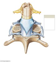
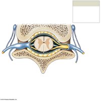
Three specialized membranes protect the spinal cord and carry blood supply:
Dura mater: Outermost, tough, dense collagen fibers.
Arachnoid mater: Middle, weblike membrane with subarachnoid space filled with CSF.
Pia mater: Innermost, mesh of collagen and elastic fibers, closely attached to the spinal cord.
Denticulate ligaments: Anchor pia mater to dura mater, preventing lateral movement.
Gray Matter and White Matter
Functional Organization of Gray Matter
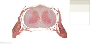
Gray matter consists of neuron cell bodies, neuroglia, and unmyelinated axons, organized into nuclei and horns:
Posterior horns: Somatic and visceral sensory nuclei.
Anterior horns: Somatic motor nuclei.
Lateral horns: Visceral motor nuclei (thoracic/lumbar).
Gray commissures: Axons crossing from one side to the other.
Functional Organization of White Matter
White matter contains myelinated and unmyelinated axons, organized into columns and tracts:
Posterior, anterior, and lateral white columns: Regions of white matter.
Tracts: Bundles of axons relaying the same type of information in the same direction.
Ascending tracts: Sensory information to the brain.
Descending tracts: Motor commands from the brain.
Spinal Nerves and Plexuses
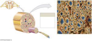
Connective Tissue Layers of Spinal Nerves
Epineurium: Outermost, dense collagen fibers.
Perineurium: Middle, surrounds fascicles (axon bundles).
Endoneurium: Innermost, surrounds individual axons.
Peripheral Distribution and Dermatomes
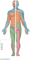
Dermatome: Region of skin supplied by a single pair of spinal nerves.
Peripheral neuropathies: Regional loss of neural function, often affecting dermatomes.
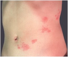
Shingles: Viral infection causing rash along dermatomes.
Nerve Plexuses
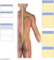
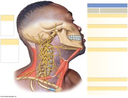
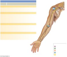
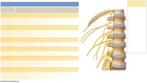
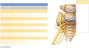
Nerve plexuses are complex networks formed by the anterior rami of adjacent spinal nerves, allowing multiple nerves to supply the same structures. The four major plexuses are:
Cervical plexus (C1–C5): Innervates neck and diaphragm (phrenic nerve).
Brachial plexus (C5–T1): Innervates pectoral girdle and upper limb.
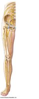
Lumbar plexus (T12–L4): Innervates pelvic girdle and lower limb.
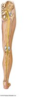
Sacral plexus (L4–S4): Innervates pelvic girdle and lower limb.
Major Nerves of the Plexuses
Cervical plexus: Phrenic, lesser occipital, great auricular, transverse cervical, supraclavicular nerves.
Brachial plexus: Musculocutaneous, median, ulnar, axillary, radial nerves.
Lumbar plexus: Iliohypogastric, ilio-inguinal, femoral, obturator, genitofemoral, lateral femoral cutaneous nerves.
Sacral plexus: Sciatic, superior/inferior gluteal, pudendal, posterior femoral cutaneous nerves.
Sensory Innervation of the Hand and Foot
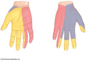
Hand: Ulnar, median, and radial nerves supply different regions; median nerve passes through carpal tunnel (carpal tunnel syndrome).
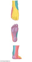
Foot: Saphenous, sural, fibular, and tibial nerves supply the foot and ankle.
Neuronal Pools and Neural Circuits
Types of Neurons and Pools
Sensory neurons: Bring information into CNS.
Motor neurons: Send commands from CNS to effectors.
Interneurons: Interpret, plan, and coordinate signals within CNS.
Neuronal pools: Functional groups of interconnected interneurons.
Patterns of Neural Circuits
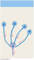
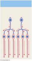
Divergence: One neuron spreads information to many (common in sensory pathways).
Convergence: Many neurons synapse on a single neuron (e.g., control of diaphragm).
Serial processing: Sequential information flow (e.g., pain signals).
Parallel processing: Simultaneous processing by several pools (e.g., response to stepping on a bee).
Reverberation: Positive feedback loops maintain activity (e.g., breathing, muscle coordination).
Reflexes and Reflex Arcs
Neural Reflexes and Reflex Arc Components
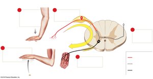
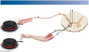
Reflexes are rapid, automatic responses to specific stimuli, forming the basic building blocks of neural function. A reflex arc consists of five components:
Sensory receptor
Sensory neuron
Information processing in CNS
Motor neuron
Effector (muscle or gland)
Classification of Reflexes
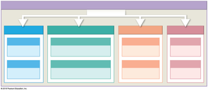
Development: Innate (inborn) or acquired (learned).
Response: Somatic (skeletal muscle) or visceral (autonomic, smooth/cardiac muscle, glands).
Complexity: Monosynaptic (one synapse) or polysynaptic (multiple synapses).
Processing site: Spinal or cranial.
Spinal Reflexes
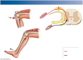
Monosynaptic Reflexes: The Stretch Reflex
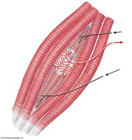
The stretch reflex is a monosynaptic reflex that regulates skeletal muscle length. It is very rapid and important for posture and balance.
Stimulus: Muscle stretching.
Receptor: Muscle spindle (intrafusal fibers).
Response: Reflexive contraction of the stretched muscle.
Polysynaptic Reflexes
Tendon reflex: Prevents excessive tension in muscles/tendons (Golgi tendon organs).
Withdrawal reflexes: Move body part away from painful stimulus; involve reciprocal inhibition.
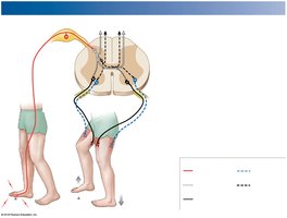
Crossed extensor reflexes: Contralateral response to maintain balance (e.g., stepping on a sharp object).
Characteristics of Polysynaptic Reflexes
Involve pools of interneurons (excitation or inhibition).
Involve multiple spinal segments.
Activate muscles in multiple areas.
Have reciprocal inhibition and reverberating circuits.
Several reflexes may cooperate for coordinated response.
Brain Control of Spinal Reflexes
Integration and Modification
Brain can facilitate, inhibit, or fine-tune spinal reflexes via descending pathways.
Reinforcement: Higher centers enhance reflexes by stimulating excitatory interneurons.
Inhibition: Higher centers suppress reflexes by stimulating inhibitory neurons or creating IPSPs.
Plantar and Babinski Reflexes
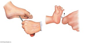
Plantar reflex: Normal in adults; stroking the sole causes toe-curling.
Babinski reflex: Normal in infants; dorsiflexion of the big toe. In adults, may indicate CNS damage.