 뒤로
The Urinary System: Tubular Reabsorption, Secretion, and Water Conservation
스터디 가이드 - 스마트 노트
 자료에 맞춘 맞춤형 노트, 핵심 정의, 예시, 맥락을 확장해 제공합니다.
The Urinary System: Tubular Reabsorption, Secretion, and Water Conservation
Overview of Renal Processes
The urinary system is responsible for filtering blood, removing waste, and regulating water and electrolyte balance. The nephron, the functional unit of the kidney, performs these tasks through glomerular filtration, tubular reabsorption, tubular secretion, and water conservation.
Glomerular Filtration: Driven by high blood pressure in glomerular capillaries, allowing water and most solutes to enter the capsular space while retaining blood cells and large proteins in the bloodstream.
Tubular Reabsorption: Returns essential substances from the filtrate back to the blood.
Tubular Secretion: Adds additional wastes and drugs from the blood into the filtrate.
Water Conservation: Adjusts water reabsorption to maintain body fluid balance.
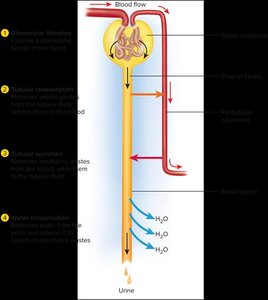
Tubular Reabsorption and Secretion
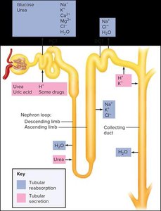
Tubular reabsorption and secretion occur throughout the nephron, including the proximal convoluted tubule (PCT), nephron loop, and distal convoluted tubule (DCT). These processes are essential for maintaining homeostasis and removing wastes.
Tubular Reabsorption: Movement of substances from the filtrate back into the blood, including electrolytes, glucose, water, and some nitrogenous wastes.
Tubular Secretion: Transfer of additional wastes, drugs, and ions from the blood into the tubular fluid.
Both processes: Occur simultaneously and are regulated by transport proteins and hormonal signals.
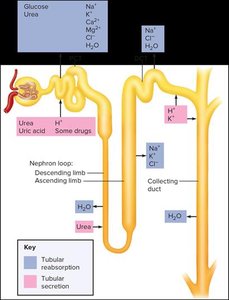
Proximal Convoluted Tubule (PCT)
The PCT reabsorbs about 65% of the filtrate and is the primary site for reabsorption of electrolytes, glucose, water, and some nitrogenous wastes. It also secretes certain wastes and drugs into the filtrate.
Electrolytes: Sodium is the most important, with transport proteins facilitating its uptake. Sodium-potassium pumps move sodium into the extracellular fluid, and chloride follows by electrical attraction. Other ions like potassium, magnesium, calcium, and phosphate are also reabsorbed.
Glucose: Co-transported with sodium.
Water: About two-thirds reabsorbed by the PCT.
Nitrogenous Wastes: Most are excreted in urine, but some are reabsorbed.
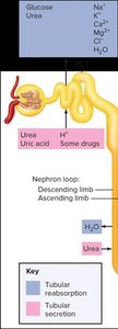
Nephron Loop (Loop of Henle)
The nephron loop generates an osmotic gradient that enables the collecting duct to reabsorb water. The thin segment is permeable to water, while the thick segment actively transports salts.
Descending Limb: Permeable to water, allowing water reabsorption.
Ascending Limb: Active transport of salts into medullary tissue, contributing to the osmotic gradient.
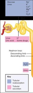
Distal Convoluted Tubule (DCT)
The DCT fine-tunes the reabsorption of water and salts, regulated by hormones such as aldosterone and natriuretic peptides.
Aldosterone: Increases sodium reabsorption when sodium is low, potassium is high, or blood pressure is low.
Natriuretic Peptides: Promote sodium and water excretion when blood pressure is high.
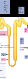
Tubular Secretion
Tubular secretion removes wastes, drugs, and excess ions from the blood and adds them to the filtrate. This process is important for acid-base balance, waste removal, and drug clearance.
Acid-Base Balance: Secretion of hydrogen and bicarbonate ions helps regulate blood pH.
Waste Removal: Urea, uric acid, and other wastes are secreted.
Drug Clearance: Many drugs are secreted into the tubular fluid for excretion.
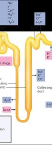
Summary of Tubular Reabsorption and Secretion
PCT: Reabsorbs 65% of filtrate.
Nephron Loop: Reabsorbs 25% of filtrate.
DCT: Reabsorbs additional sodium, chloride, and water.
Hormonal Control: Aldosterone and natriuretic peptides regulate reabsorption.
Secretion: Drugs, wastes, and other solutes are secreted into tubular fluid.
Collecting Duct and Water Conservation
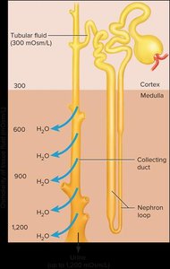
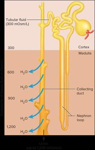
The collecting duct receives tubular fluid from multiple nephrons and is responsible for reabsorbing water and concentrating urine. The osmolarity of the surrounding tissue and the permeability of the collecting duct determine how much water is reabsorbed.
Osmolarity: The medullary portion of the collecting duct is more permeable to water, allowing water to be reabsorbed into the salty medullary tissue.
Countercurrent Multiplier: The nephron loop continuously recaptures salt and returns it to the deep medullary tissue, maintaining the osmotic gradient.
Control of Water Loss
The concentration of urine depends on the body's hydration state. Antidiuretic hormone (ADH) plays a key role in regulating water reabsorption in the collecting duct.
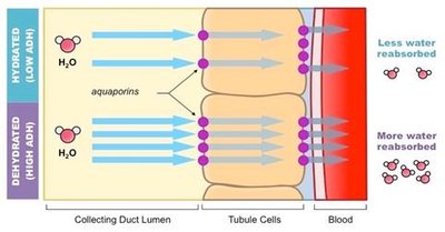
Dehydration: Increased blood osmolarity stimulates ADH release, causing collecting duct cells to produce aquaporins and reabsorb more water, resulting in concentrated urine.
Hydration: Low blood osmolarity inhibits ADH secretion, reducing aquaporin production and water reabsorption, resulting in dilute urine.
Summary of Water Conservation
Collecting Duct: Adjusts water reabsorption based on the body's needs.
ADH: Increases water reabsorption when dehydrated; decreases when hydrated.
Osmosis: Water moves from the collecting duct into the salty medulla.
Countercurrent Multiplier: Maintains medullary osmolarity for water conservation.
Voiding Urine (Micturition)
Urine is transported from the kidneys to the bladder via the ureters. Micturition, or urination, is initiated by the micturition reflex, which is triggered by stretch receptors in the bladder wall.
Micturition Reflex: Filling of the bladder excites stretch receptors, sending signals to the micturition center in the pons.
Integration: The pons can integrate information from the cerebral cortex, allowing voluntary control over urination.
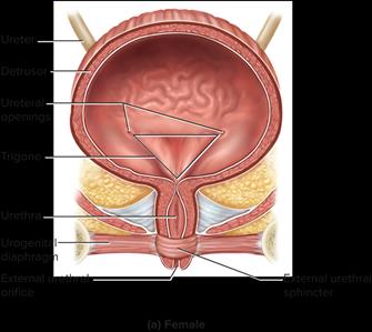
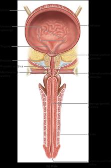
Bladder Anatomy: Includes the detrusor muscle, urethra, and sphincters.
Key Terms and Concepts
Glomerular Filtration Rate (GFR): The rate at which blood is filtered in the glomerulus; maintained by neural, hormonal, and local mechanisms.
Aldosterone: Hormone that increases sodium and water reabsorption.
Antidiuretic Hormone (ADH): Hormone that increases water reabsorption in the collecting duct.
Countercurrent Multiplier: Mechanism in the nephron loop that maintains medullary osmolarity.
Micturition: The process of voiding urine from the bladder.
Relevant Equations
Osmosis: Water movement is driven by osmotic gradients.
Glomerular Filtration Rate: where is the filtration coefficient, is glomerular hydrostatic pressure, is capsular hydrostatic pressure, and is glomerular oncotic pressure.
Example
When a person is dehydrated, blood osmolarity increases, stimulating ADH release. ADH causes the collecting duct to produce aquaporins, increasing water reabsorption and resulting in concentrated urine.
Additional info: The notes have been expanded to include definitions, examples, and relevant equations for clarity and completeness.
