 뒤로
The Visual System: Anatomy & Physiology Study Notes
스터디 가이드 - 스마트 노트
 자료에 맞춘 맞춤형 노트, 핵심 정의, 예시, 맥락을 확장해 제공합니다.
The Visual System: Structure and Function
Overview of Sensory Systems
Sensory systems are specialized networks that allow the brain to detect and interpret environmental stimuli. These systems are essential for both external perception and internal regulation.
Sensory Stimulus: A form of energy (mechanical, chemical, thermal, or light) capable of exciting a receptor.
Sensory Receptors: Specialized cells that transduce environmental energy into neural signals.
Sensation vs. Perception: Sensation is the detection of environmental stimuli; perception is the conscious experience of these stimuli, shaped by brain connections and structures.
Law of Specific Nerve Energies: Each receptor responds selectively to one form of energy, defining the modality of sensation.
Topographic Mapping in Sensory Systems
Sensory systems are organized topographically, meaning spatial relationships in the environment are preserved in neural representations.
Retinotopic: Visual system mapping
Tonotopic: Auditory system mapping
Somatotopic: Somatosensory system mapping
Chemotopic: Olfactory system mapping
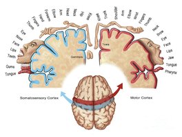
Sensory Resolution
The resolution of a sensory system depends on the density of receptors, the number of afferent connections, and the size of the primary cortical area dedicated to that sense.
High receptor density: Higher resolution
Low receptor density: Lower resolution

Receptor Potential and Rate Law
Receptor potential is the electrical change in a neuron as it transduces environmental energy. The rate law describes how stimulus intensity affects the rate of action potentials in sensory neurons.
Stimulus intensity: Determines amplitude of receptor potential
Rate of action potentials: Encodes stimulus strength
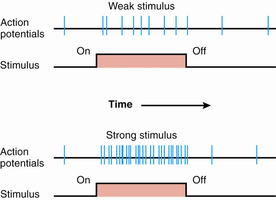
Anatomy of the Eye and Retina
Structure of the Eye
The eye is a complex organ designed to admit, focus, and transduce light.
Pupil: Admits light
Lens: Focuses light
Retina: Contains photoreceptors that transduce light
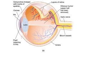
Visual Field Mapping
The visual field is mapped onto the retina, with central vision focused on the fovea and peripheral vision on the outer retina.
Retinal Flow of Information
Photoreceptors detect light and transmit signals to bipolar cells, which relay information to ganglion cells. Ganglion cells send signals to the brain via the optic nerve.
Bipolar cells: Connect photoreceptors to ganglion cells
Ganglion cells: Project to the brain
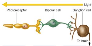
Types of Photoreceptors: Rods and Cones
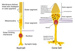
The retina contains two main types of photoreceptors, each specialized for different aspects of vision.
Rods: Sensitive to dim light, not color; more numerous in the periphery
Cones: Sensitive to color and high resolution; densely packed in the fovea
Cones | Rods |
|---|---|
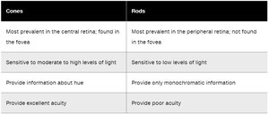
Most prevalent in central retina, found in fovea | Most prevalent in peripheral retina, not found in fovea |
Sensitive to moderate/high light | Sensitive to low light |
Provide information about hue | Provide only monochromatic information |
Provide excellent acuity | Provide poor acuity |
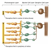
Fovea and Peripheral Retina
The fovea is the central region of the retina with a high density of cones, providing sharp, detailed vision. The periphery contains more rods, supporting vision in low light.
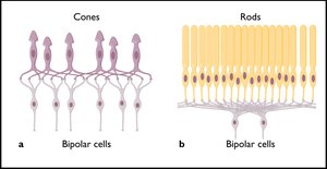
Convergence and Visual Acuity
Convergence refers to the number of photoreceptors synapsing onto a single bipolar cell. High convergence (rods) amplifies dim light but reduces resolution; low convergence (cones) enhances acuity.
Horizontal and Amacrine Cells
Horizontal and amacrine cells connect adjacent photoreceptors and bipolar cells, facilitating lateral communication and contrast enhancement.
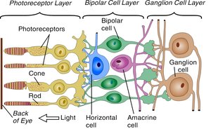
Transduction of Visual Stimuli
Phototransduction Mechanism
Light activates photopigments in rods and cones, triggering a cascade that leads to hyperpolarization and reduced glutamate release.
Rod opsin and retinal: Separate upon light exposure
Hyperpolarization: Decreases glutamate release
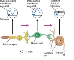
Bipolar Cell Responses
Bipolar cells are classified as ON-center or OFF-center, responding differently to glutamate. This enhances contrast and edge detection.
ON-center: Hyperpolarized by glutamate
OFF-center: Depolarized by glutamate
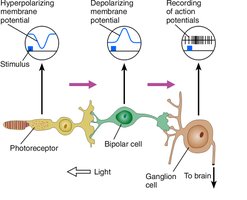
Pathway of Visual Information to the Brain
Optic Nerve and Optic Chiasm
Axons from ganglion cells form the optic nerve, which partially crosses at the optic chiasm. Nasal fibers cross to the contralateral side.
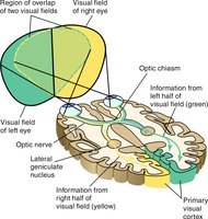
Lateral Geniculate Nucleus (LGN)
The LGN is a six-layered structure in the thalamus that processes visual information. Layers differ by input (contralateral vs. ipsilateral eye) and function (movement, color, detail).
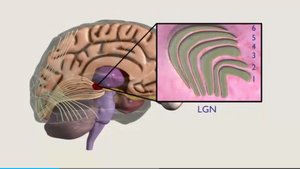
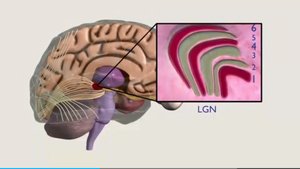
Layer | Type of Information |
|---|---|
Magnocellular | Movement |
Parvocellular | Color and Detail |
Koniocellular | Color (between layers) |
Primary Visual Cortex (V1/Striate Cortex)
The primary visual cortex is organized into six layers, with layer 4c receiving LGN input. Neurons respond to specific features such as orientation, movement, and spatial patterns.
Modular Organization and Feature Processing
V1 is organized into modules that process orientation, ocular dominance, and color. These modules communicate to build a feature-specific representation of the visual field.
Secondary Visual Cortex (V2)
Information from V1 modules is combined in V2, allowing for object and scene perception. Processing is hierarchical, with layers close to V1 handling modular information.
Dorsal and Ventral Streams
The visual system splits into two streams:
Dorsal stream: "Where" pathway, projects to parietal cortex for spatial analysis
Ventral stream: "What" pathway, projects to temporal cortex for object recognition
Summary Table: Visual System Pathway
Step | Structure | Function |
|---|---|---|
1 | Photoreceptors | Transduce light |
2 | Bipolar cells | Relay signals |
3 | Ganglion cells | Send signals to brain |
4 | Optic nerve/chiasm | Transmit and cross signals |
5 | LGN | Process and relay information |
6 | V1 (Striate cortex) | Feature analysis |
7 | V2 (Visual association cortex) | Object and scene perception |
Additional info: Academic context was added to clarify the function and organization of the visual system, including the role of topographic mapping, cell types, and pathways.
