 뒤로
뒤로Tissue: The Living Fabric – Epithelial Tissue Study Guide
스터디 가이드 - 스마트 노트
 자료에 맞춘 맞춤형 노트, 핵심 정의, 예시, 맥락을 확장해 제공합니다.
Tissues and Histology
Definition and Importance
Tissues are groups of cells similar in structure and function, forming the basic building blocks of organs. Histology is the study of tissues, essential for understanding organ structure and function. Most organs contain several tissue types, and their arrangement determines the organ’s capabilities.
Epithelial tissue: Lining, covering, and glandular tissue
Connective tissue: Supportive and nutritive
Muscular tissue: Contracts to produce movement
Nervous tissue: Integration and control
Epithelial Tissue
Characteristics of Epithelial Tissue
Epithelial tissue consists of closely packed cells forming continuous sheets, which may be single or multiple layers. The apical layer is exposed to the body’s exterior or internal organ cavities, and may have modifications such as microvilli or cilia. The basal layer is attached to a basement membrane composed of a basal lamina made of glycoprotein.
Functions: Protection, absorption, filtration, secretion
Avascular: No blood supply; relies on diffusion from capillaries
Innervated: Supplied by nerve fibers
High regeneration: Well-nourished cells can reproduce rapidly
Cell junctions: Tight junctions, desmosomes, gap junctions

Naming and Classification of Epithelium
Epithelial tissues are named based on cell shape and arrangement:
Shapes: Squamous (thin, flat), Cuboidal (cube-shaped), Columnar (column-shaped)
Arrangements: Simple (single layer), Stratified (multiple layers), Pseudostratified (appears stratified, but is not), Transitional (stretchable), Ciliated (possess cilia)
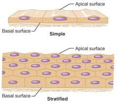
How to Name Epithelial Tissue
First name: Number of layers (simple or stratified)
Second name: Shape of cells (squamous, cuboidal, columnar)
Types of Epithelial Tissue
Simple Squamous Epithelium
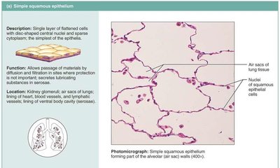
This is the thinnest tissue in the body, forming semipermeable membranes for transport. Found in alveolar membranes (lungs), capillary walls, and kidneys.
Function: Allows transport of gases, nutrients, wastes
Basement membrane: Important filter for substances

Endothelium and Mesothelium
Endothelium: Lines lymphatic vessels and cardiovascular system
Mesothelium: Lines serous membranes of ventral body cavity and organs
Simple Cuboidal Epithelium
Single layer of cube-shaped cells forming ducts of glands, kidney tubules, and ovary surface.
Simple Columnar Epithelium
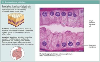
Single layer of column-shaped cells, with nuclei near the basal layer. Apical surface may have microvilli or cilia. Non-ciliated type is found in the GI tract; ciliated type in respiratory and reproductive tracts.
Microvilli: Increase surface area for absorption (small intestine)
Goblet cells: Secrete mucus for protection and lubrication
Stratified Epithelia
Contain two or more cell layers, regenerating from below. Function is protection; identified by the shape of the upper layer.
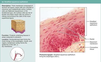
Stratified Squamous Epithelium
Most widespread stratified epithelium
Surface cells are squamous; deeper cells are cuboidal or columnar
Forms epidermis of skin and lines body openings (mouth, anus, vagina)
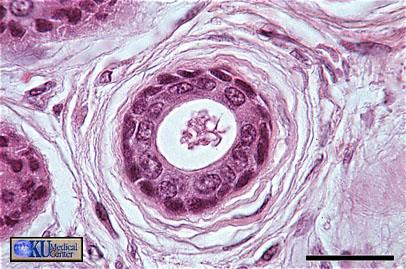
Stratified Cuboidal Epithelium
Typically two layers of cuboidal cells
Rare; found in ducts of large glands (mammary, sweat glands)
Stratified Columnar Epithelium
Limited distribution; found in pharynx, male urethra, glandular ducts
Occurs at transition areas between other epithelia
Only apical layer is columnar
Pseudostratified Ciliated Columnar Epithelium
Single layer of columnar cells of uneven height; nuclei at different levels. Ciliated type lines respiratory tract; non-ciliated found in large glands and male urethra.
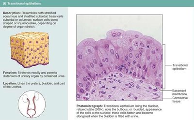
Transitional Epithelium
Highly modified stratified squamous epithelium, found in bladder, ureters, and urethra. Allows stretchability to prevent excessive pressure during urination. Basal cells are cuboidal or columnar; apical cells change shape depending on organ fullness.
Glandular Epithelium
Definition and Types
A gland consists of one or more cells producing a secretion, usually a protein in aqueous solution or lipid-rich (oil glands). Two main types:
Exocrine glands: Have ducts; secrete onto epithelial surfaces
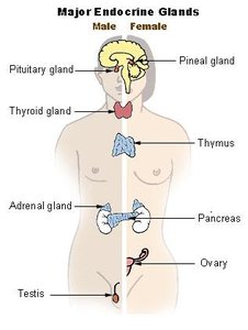
Endocrine glands: Ductless; secrete hormones directly into blood or lymph
Endocrine Glands
Ductless glands
Secrete hormones into blood or lymph
Examples: Thyroid, pituitary
Exocrine Glands
Have ducts
May be unicellular (goblet cells) or multicellular (sweat, oil glands)
Secretions empty through ducts onto surfaces
Goblet cells secrete mucin, forming mucus for protection and lubrication
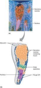
Multicellular Exocrine Glands
Complex structure with two basic parts: epithelium-derived duct and secretory unit. Supported by connective tissue, which supplies blood vessels and nerves, and forms a fibrous capsule dividing the gland into lobes.
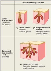
Classification of Multicellular Exocrine Glands
Based on duct type and secretory unit:
Simple glands: Unbranched ducts
Compound glands: Branched ducts
Tubular: Secretory cells form tubes
Alveolar: Secretory cells form flask-like sacs
Tubuloalveolar: Both types present
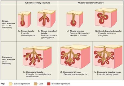

Mode of Secretion – Functional Classification
Merocrine glands: Secrete products by exocytosis; cells remain intact (e.g., pancreas, sweat, salivary glands)
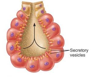
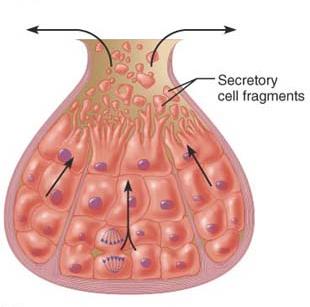
Holocrine glands: Accumulate products until rupture; secretion includes cell fragments (e.g., sebaceous glands)
Summary Table: Types of Multicellular Exocrine Glands
Type | Duct Structure | Secretory Structure | Example |
|---|---|---|---|
Simple tubular | Unbranched | Tubular | Intestinal glands |
Simple branched tubular | Unbranched | Branched tubular | Stomach (gastric) glands |
Simple alveolar | Unbranched | Alveolar | No important example in humans |
Simple branched alveolar | Unbranched | Branched alveolar | Sebaceous (oil) glands |
Compound tubular | Branched | Tubular | Duodenal glands of small intestine |
Compound alveolar | Branched | Alveolar | Mammary glands |
Compound tubuloalveolar | Branched | Tubuloalveolar | Salivary glands |
Key Terms
Epithelium: Tissue forming linings and coverings
Basement membrane: Glycoprotein-rich layer anchoring epithelium
Goblet cell: Unicellular gland secreting mucus
Merocrine: Secretion by exocytosis
Holocrine: Secretion by cell rupture
Additional info:
Transitional epithelium is unique for its ability to stretch and change shape, crucial for organs like the bladder.
Cell junctions (tight, desmosomes, gap) are essential for maintaining tissue integrity and communication.
