 뒤로
뒤로Tissues: Structure, Types, and Functions in Human Anatomy
스터디 가이드 - 스마트 노트
 자료에 맞춘 맞춤형 노트, 핵심 정의, 예시, 맥락을 확장해 제공합니다.
Tissues
Definition and Overview
Tissues are groups of cells linked together to perform specialized functions within the human body. Understanding tissue types and their characteristics is fundamental to the study of anatomy and physiology.
Definition: Groups of cells working together for a common function.
Main Functions: Protection, absorption, secretion, sensation.
Basic Tissue Types
Classification of Tissues
The human body contains four primary tissue types, each with distinct roles:
Epithelial tissue
Connective tissue
Muscle tissue
Neural (nervous) tissue
Epithelial Tissue
Characteristics of Epithelial Tissue
Epithelial tissue forms the covering or lining of internal and external surfaces. It is highly cellular, with cells closely bound together, and features a distinct apical surface exposed to the environment or internal space, and a basal surface attached to a basement membrane.
Cellularity: Numerous, tightly packed cells.
Apical Surface: Exposed to external environment or internal cavity.
Basement Membrane: Anchors epithelium to underlying tissue.
Avascular: Lacks blood vessels; nutrients diffuse from underlying tissues.
Regeneration: High mitotic activity allows rapid repair.
Functions of Epithelial Tissue
Protection: Covers surfaces to shield underlying tissues.
Absorption: Uptake of substances (e.g., nutrients in intestines).
Secretion: Production and release of substances (e.g., mucus, hormones).
Sensation: Contains sensory receptors.
Types of Epithelial Tissue
Epithelial tissues are classified based on the number of cell layers and cell shape:
Simple epithelium: Single layer of cells.
Stratified epithelium: Multiple layers of cells.
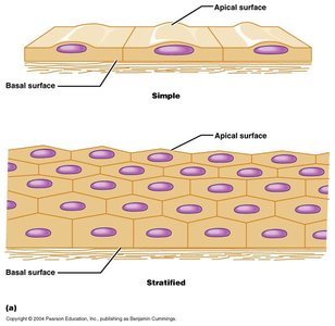
Squamous: Flat cells; can be simple or stratified.
Cuboidal: Cube-shaped; can be simple, stratified, or transitional.
Columnar: Tall, column-like; can be simple, pseudostratified, or stratified.
Glands
Glands are specialized epithelial structures that produce and secrete substances. They are classified as endocrine or exocrine:
Endocrine glands: Ductless; release hormones directly into the bloodstream.
Exocrine glands: Have ducts; release secretions onto surfaces or into cavities.
Exocrine Gland Secretions
Exocrine glands are further classified by their mode of secretion:
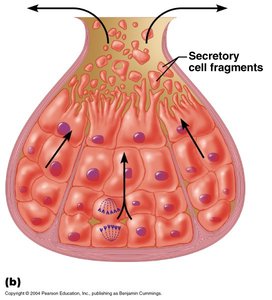
Merocrine: Secrete products via exocytosis; cells remain intact. Examples: salivary glands, pancreas.
Apocrine: Portion of cell pinches off with secretion; historically associated with mammary glands.
Holocrine: Entire cell disintegrates to release product; seen in sebaceous (oil) glands.
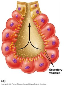
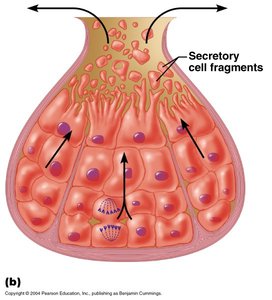
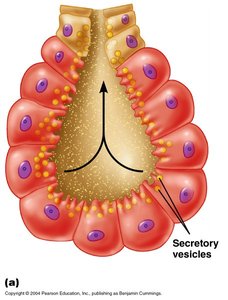
Exocrine Gland Structure
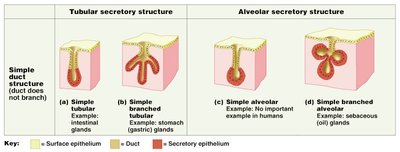
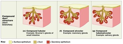
Exocrine glands can be unicellular (single cell, e.g., goblet cell) or multicellular. Multicellular glands are classified by their duct and secretory unit structure:
Simple glands: Unbranched duct.
Compound glands: Branched duct.
Tubular: Tube-shaped secretory unit.
Acinar (alveolar): Round, sac-like secretory unit.
Tubuloacinar: Combination of tubular and acinar units.
Connective Tissue
Features of Connective Tissue
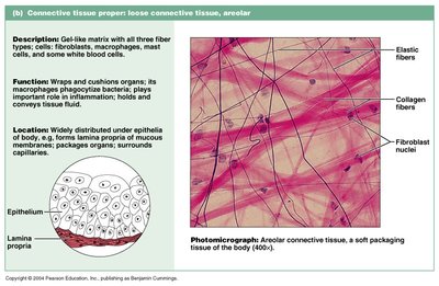
Connective tissue supports, binds, and protects other tissues and organs. It is characterized by sparse cells and abundant extracellular matrix composed of protein fibers and ground substance.
Specialized cells: Fibroblasts, macrophages, adipocytes, melanocytes, mast cells, white blood cells.
Matrix: Non-living material; includes protein fibers (collagen, elastic, reticular) and ground substance (water, proteins, hyaluronic acid).
Vascularity: Most connective tissues are vascular, except cartilage, tendons, and ligaments.
Limited mitotic activity: Slower repair compared to epithelial tissue.
Functions of Connective Tissue
Framework: Structural support for the body.
Transport: Blood and lymph are fluid connective tissues.
Protection: Physical and immunological defense.
Support: Maintains shape and structure.
Energy storage: Adipose tissue stores fat.
Defense: Immune cells present in connective tissue.
Types of Connective Tissue
Connective tissue proper: Loose (areolar, adipose, reticular) and dense (regular, irregular, elastic).
Fluid connective tissue: Blood, lymph.
Supporting connective tissue: Cartilage, bone.
Connective Tissue Fibers
Collagen: Strong, provides tensile strength.
Reticular: Forms supportive networks.
Elastic: Provides elasticity and resilience.
Connective Tissue Cells
Fibroblasts: Produce fibers and ground substance.
Macrophages: Phagocytic cells; defense.
White blood cells: Immune response.
Adipocytes: Store fat.
Melanocytes: Produce pigment.
Mast cells: Release histamine; involved in inflammation.
Marfan's Syndrome
Marfan's syndrome is a genetic disorder affecting connective tissue due to abnormal glycoprotein fibrillin, which forms elastic fibers. It impacts all connective tissues, leading to long limbs, weakened blood vessels, and risk of aneurysms.
Membranes
Types of Membranes
Membranes are sheets of tissue that cover or line body surfaces. They are classified as epithelial or synovial membranes.
Epithelial membranes: Composed of an upper epithelial layer and a lower connective tissue layer.
Synovial membranes: Composed only of connective tissue; found in joints.
Epithelial Membranes
Mucosa: Lines cavities open to the outside (digestive, respiratory, urinogenital); contains mucus-producing epithelial cells and a connective layer called lamina propria.
Serosa: Lines cavities not open to the outside (abdomen, thorax); produces serous fluid; consists of parietal and visceral layers.
Cutaneous: Skin; protects the body.
Synovial Membranes
Structure: Loose connective tissue; no epithelial layer.
Function: Lines cavities of freely movable joints; secretes synovial fluid for lubrication.
Skin
Functions of Skin
The skin is the largest organ of the body, providing multiple protective and regulatory functions.
Protection: Shields from infection, water loss, UV radiation.
Insulation: Maintains body temperature.
Vitamin D production: Synthesized in response to sunlight.
Sensation: Contains sensory receptors.
Waste removal: Excretes waste products via sweat.
Skin Layers
Epidermis: Stratified squamous epithelium.
Dermis: Loose and dense connective tissue.
Hypodermis (subcutaneous): Adipose tissue.
Summary Table: Types of Epithelial and Connective Tissues
Type | Structure | Function | Example |
|---|---|---|---|
Simple Epithelium | Single layer | Absorption, filtration | Intestinal lining |
Stratified Epithelium | Multiple layers | Protection | Skin epidermis |
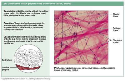
Areolar Connective | Loose matrix | Wraps, cushions organs | Under epithelia |
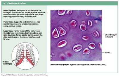
Hyaline Cartilage | Firm matrix | Support, reinforcement | Trachea, joints |
Adipose Tissue | Fat cells | Energy storage | Subcutaneous layer |
Key Equations
While tissue structure is not typically described by equations, the following formula is relevant for understanding cell division in epithelial tissue:
Mitotic Index:
Additional info: Academic context was added to expand brief points and clarify tissue types, functions, and examples.
