 뒤로
Transmembrane Potential and Action Potentials in Neurons
스터디 가이드 - 스마트 노트
 자료에 맞춘 맞춤형 노트, 핵심 정의, 예시, 맥락을 확장해 제공합니다.
Transmembrane Potential
Overview of Neuronal Electrical Properties
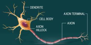
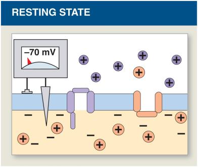
Neurons are specialized cells that generate and propagate electrical signals, essential for communication within the nervous system. The transmembrane potential is the voltage difference across the cell membrane, resulting from the distribution of ions.
Electrochemical Gradient: The combined effect of electrical and chemical gradients drives ion movement across membranes.
Membrane Channels: Ion channels on dendrites and cell bodies regulate permeability and electrical signaling.
Resting Potential: The typical resting membrane potential is approximately –70 mV, maintained by ion gradients.
Initiation and Generation of Action Potentials
Action Potential Basics
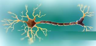
An action potential is a rapid, propagated change in transmembrane potential that affects the entire excitable membrane. It links graded potentials at the cell body with activities at the postsynaptic cell.
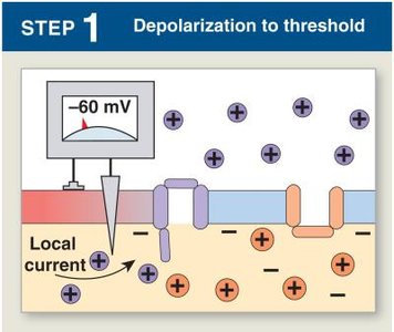
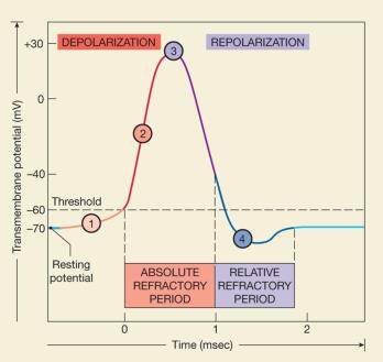
Threshold: A graded depolarization at the axon hillock must be large enough (10–15 mV) to change the resting potential from –70 mV to the threshold level (–60 to –55 mV).
All-or-None Principle: If the stimulus exceeds the threshold, the action potential is triggered and always has the same magnitude.
Steps in Action Potential Generation
The generation of an action potential involves four distinct steps:
Depolarization to Threshold: Local currents depolarize the membrane to the threshold level.
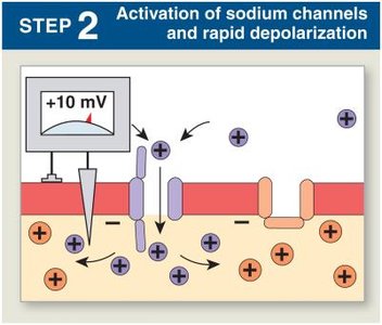
Activation of Sodium Channels: Voltage-gated Na+ channels open, causing rapid depolarization as Na+ ions rush into the cell. The inner membrane becomes positive.
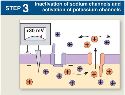
Inactivation of Sodium Channels and Activation of Potassium Channels: At +30 mV, Na+ channels close (inactivation gates), and K+ channels open, initiating repolarization.
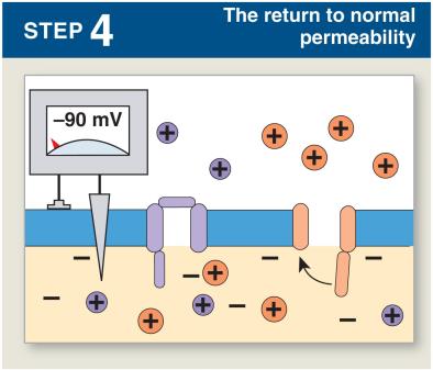
Return to Normal Permeability: K+ channels close as the membrane returns to resting potential, often hyperpolarizing to –90 mV before stabilizing.
The Refractory Period
The refractory period is the time during which the membrane will not respond normally to additional stimuli:
Absolute Refractory Period: Na+ channels are open or inactivated; no action potential can be generated.
Relative Refractory Period: Membrane potential is almost normal; a very large stimulus may initiate an action potential.
Sodium–Potassium Exchange Pump
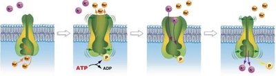
The sodium–potassium pump actively transports 3 Na+ out and 2 K+ into the cell, powered by ATP. This maintains the ion gradients and resting potential.
Balances passive diffusion: Ensures stable resting potential.
ATP dependence: Without ATP, neurons cease to function.
Propagation of Action Potentials
Continuous Propagation (Unmyelinated Axons)
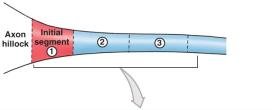
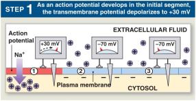
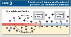
In unmyelinated axons, action potentials propagate by affecting one segment at a time. The cycle repeats as local currents depolarize adjacent segments.
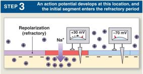
Action potential develops in segment 1, depolarizing to +30 mV.
Local current depolarizes segment 2 to threshold, generating an action potential.
Segment 1 enters refractory period.
Cycle repeats, action potential travels in one direction (1 m/sec).
Saltatory Propagation (Myelinated Axons)
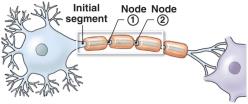
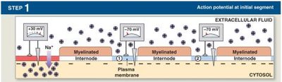
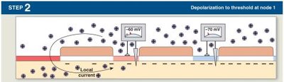
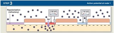
In myelinated axons, action potentials propagate faster and with less energy. Myelin insulates the axon, causing local currents to "jump" from node to node (nodes of Ranvier).
Depolarization occurs only at nodes.
Speed: Saltatory propagation is significantly faster than continuous propagation.
Axon Diameter and Speed
Influence of Axon Diameter
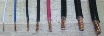
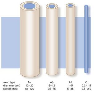
The speed of action potential propagation is influenced by axon diameter. Larger diameters offer less resistance to ion movement, resulting in faster conduction.
Critical information: Vision, balance, and urgent motor commands are carried by large-diameter, myelinated axons.
Classification of Peripheral Nerve Fibers
Peripheral nerve fibers are classified by diameter, myelination, and speed:
Type | Diameter | Myelination | Speed | Function |
|---|---|---|---|---|
A (α, β, γ, δ) | Large | Myelinated | High (up to 120 m/sec) | Rapid information, motor control |
B | Medium | Some myelinated | Intermediate (18 m/sec) | Visceral afferent, ANS preganglionic |
C | Small | Unmyelinated | Low (1 m/sec) | Slow sensory, pain, involuntary muscle |
Synapses
Electrical Synapses
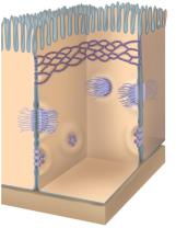
Electrical synapses involve direct physical contact between cells via gap junctions, allowing ions to pass and producing continuous local current and action potential propagation.
Chemical Synapses
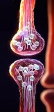
Chemical synapses are the most common, where neurotransmitters transmit signals across a gap. The effect depends on the amount of neurotransmitter released and the sensitivity of the postsynaptic cell.
Neurotransmitters
Neurotransmitters are classified as excitatory or inhibitory:
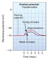
Excitatory: Cause depolarization and promote action potentials.
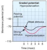
Inhibitory: Cause hyperpolarization and suppress action potentials.
Cholinergic Synapses
Cholinergic synapses release acetylcholine (ACh), which is widely distributed in the nervous system. Events include ACh release, binding, and breakdown by acetylcholinesterase (AChE).

Information Processing in Neurons
Postsynaptic Potentials
Postsynaptic potentials are graded responses to neurotransmitters:
EPSP (Excitatory Postsynaptic Potential): Graded depolarization.
IPSP (Inhibitory Postsynaptic Potential): Graded hyperpolarization.
Summation
EPSPs and IPSPs combine through summation:
Temporal Summation: Multiple signals over time.
Spatial Summation: Multiple signals from different locations.
Key Equations
Resting Membrane Potential
The resting membrane potential is determined by the Nernst equation:
Sodium–Potassium Pump
For each cycle:
Summary Table: Action Potential Steps
Step | Event | Membrane Potential |
|---|---|---|
1 | Depolarization to threshold | -60 mV |
2 | Activation of Na+ channels | +10 mV |
3 | Inactivation of Na+ channels, activation of K+ channels | +30 mV |
4 | Return to normal permeability | -90 mV |
Additional info: Academic context was added to clarify the steps of action potential generation, propagation, and synaptic transmission, as well as to provide definitions and examples for key terms.
