 뒤로
Unit 1 Study Guide: Endocrine System, Heart, and Blood Vessels
스터디 가이드 - 스마트 노트
 자료에 맞춘 맞춤형 노트, 핵심 정의, 예시, 맥락을 확장해 제공합니다.
Endocrine System
Hormones of the Adrenal Gland and Their Functions
The adrenal gland produces several hormones essential for stress response, metabolism, and electrolyte balance.
Cortisol: Regulates metabolism, immune response, and stress adaptation.
Aldosterone: Controls sodium and potassium balance, affecting blood pressure.
Epinephrine & Norepinephrine: Mediate the 'fight or flight' response, increasing heart rate and blood flow to muscles.
Anatomical and Physiological Connections of the Hypothalamus and Pituitary Glands
The hypothalamus and pituitary glands form a central regulatory axis for endocrine function.
Anatomical Connection: The hypothalamus is connected to the pituitary via the infundibulum.
Physiological Connection: The hypothalamus releases hormones that control the anterior pituitary (e.g., TRH, CRH), while the posterior pituitary stores and releases hormones produced by the hypothalamus (e.g., ADH, oxytocin).
Functional Classifications of Hormones
Hormones are classified based on their chemical structure and mechanism of action.
Amino acid-based hormones: Include peptides and proteins (e.g., insulin, growth hormone).
Steroid hormones: Derived from cholesterol (e.g., cortisol, aldosterone).
Mechanism: Hormones may act via cell surface receptors (peptides) or intracellular receptors (steroids).
Hormones of Calcium Homeostasis
Calcium levels are regulated by three main hormones.
Parathyroid hormone (PTH): Increases blood calcium by stimulating bone resorption and kidney reabsorption.
Calcitonin: Lowers blood calcium by inhibiting bone resorption.
Vitamin D (Calcitriol): Enhances intestinal absorption of calcium.
Thymus Gland
The thymus is involved in immune system development, especially during childhood.
Function: Maturation of T-lymphocytes (T-cells).
Hormones: Thymosin, which promotes T-cell development.
Homeostasis of Blood Sugar
Blood glucose is regulated by pancreatic hormones.
Insulin: Lowers blood glucose by promoting uptake into cells.
Glucagon: Raises blood glucose by stimulating glycogen breakdown.
Cardiovascular System: The Heart
The Heart Anatomy and Conduction System
The heart is a muscular organ with four chambers and a specialized conduction system for coordinated contraction.
Chambers: Right atrium, right ventricle, left atrium, left ventricle.
Conduction System: Includes the SA node, AV node, bundle of His, bundle branches, and Purkinje fibers.
Blood Flow in the Chambers of the Heart
Blood flows through the heart in a specific sequence, ensuring oxygenation and circulation.
Right atrium → right ventricle → pulmonary artery → lungs → left atrium → left ventricle → aorta → body.
Anatomy of the Walls of the Heart
The heart wall consists of three layers.
Epicardium: Outer layer.
Myocardium: Muscular middle layer responsible for contraction.
Endocardium: Inner lining.
Coronary Cardiac Circuit Anatomy
The coronary arteries supply blood to the heart muscle.
Right and left coronary arteries: Branch from the aorta.
Cardiac veins: Drain blood into the coronary sinus.
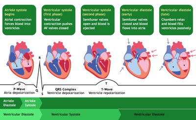
Cardiac Cycle Events – 4 Phases
The cardiac cycle consists of four main phases, each corresponding to electrical and mechanical events.
Atrial systole: Atria contract, pushing blood into ventricles.
Ventricular systole (first phase): Ventricles contract, AV valves close.
Ventricular systole (second phase): Semilunar valves open, blood is ejected.
Ventricular diastole: Ventricles relax, semilunar valves close, blood flows into atria and ventricles passively.
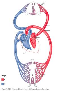
Pulmonary and Systemic Blood Circulation
The heart pumps blood through two main circuits: pulmonary and systemic.
Pulmonary circuit: Right side of heart pumps deoxygenated blood to lungs.
Systemic circuit: Left side of heart pumps oxygenated blood to the body.
Cardiac Output Law
Cardiac output is the volume of blood pumped by the heart per minute.
Formula:
Normal values: 4-8 L/min in adults.
Stroke Volume Laws
Stroke volume is determined by preload, afterload, and contractility.
Preload: Degree of stretch of cardiac muscle before contraction.
Afterload: Resistance the heart must overcome to eject blood.
Contractility: Strength of contraction.
ECG: All Peaks, Segments, and Intervals
An electrocardiogram (ECG) records the electrical activity of the heart.
P-wave: Atrial depolarization.
QRS complex: Ventricular depolarization.
T-wave: Ventricular repolarization.
PR interval: Time from atrial to ventricular depolarization.
ST segment: Time between ventricular depolarization and repolarization.
Sinus Rhythm
Sinus rhythm is the normal rhythm of the heart, initiated by the SA node.
Regular rate (60-100 bpm), consistent P-QRS-T pattern.
Sounds of the Heart
Heart sounds are produced by valve closures.
S1: Closure of AV valves (beginning of systole).
S2: Closure of semilunar valves (end of systole).
Athlete ESV, EDV
End-systolic volume (ESV) and end-diastolic volume (EDV) are measures of ventricular volume.
EDV: Volume of blood in ventricle at end of diastole.
ESV: Volume of blood remaining after systole.
Athletes often have higher EDV and lower ESV due to increased cardiac efficiency.
Blood Vessels and Circulation
Anatomy of the Blood Vessels
Blood vessels include arteries, veins, and capillaries.
Arteries: Carry blood away from the heart.
Veins: Carry blood toward the heart.
Capillaries: Sites of exchange between blood and tissues.
Blood Pressure Forces Components
Blood pressure is determined by cardiac output, blood volume, and resistance.
Systolic pressure: Pressure during ventricular contraction.
Diastolic pressure: Pressure during ventricular relaxation.
Blood Pressure Modifying Factors
Several factors influence blood pressure.
Neural: SNS and PSNS activity.
Hormonal: Epinephrine, angiotensin II.
Renal: Regulation of blood volume.
Blood Pressure Variation Throughout the Body
Blood pressure is highest in arteries and decreases through capillaries and veins.
Arterial pressure > capillary pressure > venous pressure.
Role of SNS and PSNS in BP Regulation
The autonomic nervous system regulates blood pressure.
SNS: Increases heart rate and vasoconstriction.
PSNS: Decreases heart rate.
Mean Arterial Pressure
Mean arterial pressure (MAP) is the average pressure in arteries during one cardiac cycle.
Formula:
Types of Capillaries
Capillaries are classified by permeability.
Continuous: Least permeable, most common.
Fenestrated: Have pores, found in kidneys and intestines.
Sinusoidal: Most permeable, found in liver and spleen.
Mechanisms of Blood Pressure Regulation
Blood pressure is regulated by neural, hormonal, and renal mechanisms.
Baroreceptors: Detect changes in pressure.
Renin-angiotensin system: Increases blood volume and pressure.
Capillaries Net Filtration Factors
Net filtration in capillaries depends on hydrostatic and osmotic pressures.
Formula:
Upper Limb Arteries
Major arteries of the upper limb include:
Brachial artery, radial artery, ulnar artery, subclavian artery.
Major Veins
Major veins include:
Superior and inferior vena cava, jugular veins, femoral vein.
Case Studies
Case Study 1: Thyroid Gland Disorders
Thyroid disorders involve abnormal hormone levels and feedback mechanisms.
Hormones: T3, T4, TSH.
Feedback: High T3/T4 suppress TSH; low T3/T4 increase TSH.
Case Study 2: Heart Interventricular Defect
An interventricular wall defect affects stroke volume and EKG.
Stroke Volume: May decrease due to inefficient pumping.
EKG: May show abnormal QRS complex.
Additional info: Academic context was added to expand brief points and ensure completeness.
