 뒤로
Urinary System and Fluid & Acid-Base Balance: Study Notes
스터디 가이드 - 스마트 노트
 자료에 맞춘 맞춤형 노트, 핵심 정의, 예시, 맥락을 확장해 제공합니다.
Urinary System Anatomy & Physiology
Overview of the Urinary System
The urinary system is essential for maintaining homeostasis by regulating the composition and volume of blood, removing waste products, and controlling blood pressure and pH. It consists of specialized cells, tissues, and organs that work together to filter blood and produce urine.
Cells: Includes various epithelial tissue, transitional epithelia, thin squamous, connective tissue, and smooth muscle cells. hollow spaces insides
Tissues: Primarily epithelial (lining tubules) and smooth muscle (in walls of organs), peristaltic contraction
Organs: Kidneys, Ureters, Urinary bladder, Urethra.
Functions: Regulate plasma concentrations (osmolarity, blood pH), blood volume & pressure, produce hormones, regulate glucose, and excrete wastes.
Kidney Structure
The kidneys are the primary organs of the urinary system, responsible for filtering blood and forming urine. Each kidney contains millions of nephrons, the functional units.
Nephrons: Filter blood, reabsorb useful substances, and secrete wastes.
Collecting duct: Collects urine from nephrons.
Papillary duct: urine dumped into the duct
Renal papilla: where the papillary duct empties into
Calyx (major & minor): Cuplike structures that catch the urine. Fluid flows first into several minor calyces (each wrapping around a renal papilla) which then merge into fewer, larger major calyces
Renal pelvis : A single, large funnel-shaped cavity that collects all urine draining from the major calyx
Ureter: The muscular tube that transports urine out of the kidney's renal pelvis and down to the urinary bladder for storage.
Kidney Blood Supply
Blood supply to the kidneys is crucial for filtration and urine formation. The nephron receives blood via afferent arterioles, which form the glomerulus, and then efferent arterioles, which supply peritubular capillaries or vasa recta.
Afferent arterioles: Bring blood to the glomerulus.
Glomerulus: Capillary network for filtration.
Efferent arterioles: Carry blood away from the glomerulus.
Peritubular capillaries/vasa recta: Surround nephron tubules for reabsorption and secretion.
Nephron Structure & Function
Renal Corpuscle & Tubule
The nephron consists of the renal corpuscle (site of filtration) and the renal tubule (site of reabsorption and secretion).
Renal corpuscle: Filtration Barrier. Includes glomerular capsule, arterioles, and glomerulus(intraglomerular mesangial cells).
Juxtaglomerular complex: Increase/ decrease arterial size. Secrete renin. Regulates blood pressure and filtration rate (macula densa, juxtaglomerular cells, renin, and extraglomerular mesangial cells (feedback mechanism)).
Renal tubule: Proximal convoluted tubule (PCT), nephron loop, distal convoluted tubule (DCT), collecting duct.
Cortical vs. juxtamedullary nephrons: Differ in location and function. Cortical, shorter. juxtamedullary nephrons, longer, are important for urine concentration).
EPO location? Not sure exactly where but kidney does produce EPO
Urine Formation
Urine formation involves three main processes: filtration, reabsorption, and secretion. The kidneys remove waste products such as urea, creatinine, and uric acid, while reabsorbing useful materials.
Filtration: Occurs at the glomerulus; produces filtrate.
Reabsorption: Movement of substances from filtrate back into blood.
Secretion: Addition of substances from blood into filtrate.
Urine concen/tration: Can be up to 4x blood levels.
Filtration Mechanisms
Filtration Membrane
The filtration membrane consists of glomerular endothelium (with fenestrations), dense layer, and filtration slits formed by podocyte pedicels. It allows selective passage of substances.
Glomerular blood hydrostatic pressure (GHP): Drives filtration.
Capsular hydrostatic pressure (CsHP): Opposes filtration.
Blood colloid osmotic pressure (BCOP): Opposes filtration.
Net filtration pressure (NFP): Determines overall filtration.
Glomerular filtration rate (GFR): Volume of filtrate produced per minute.
GFR Regulation
GFR is regulated by autoregulation (myogenic mechanism, tubuloglomerular feedback), hormonal regulation (RAA system, ANP & BNP), and neural regulation.
Autoregulation: Maintains stable GFR despite changes in blood pressure.
Hormonal regulation: RAA system increases GFR; ANP & BNP decrease GFR.
Neural regulation: Sympathetic stimulation can decrease GFR.
Reabsorption & Secretion
Mechanisms of Transport
Reabsorption and secretion in the nephron occur via active (primary and secondary) and passive (diffusion, osmosis) mechanisms. Some processes are obligatory (always occur), while others are facultative (regulated by hormones).
Active transport: Requires ATP; includes symporters and antiporters.
Passive transport: Includes diffusion and osmosis.
Problem of Tm: Transport maximum limits reabsorption of substances.
Proximal Convoluted Tubule (PCT)
The PCT is responsible for most reabsorption, including Na+ transport via symporters and antiporters, and passive processes like diffusion and osmosis (aquaporin-1 channels).
Na+ symporters: Move Na+ with other substances.
Na+ antiporters: Exchange Na+ for other ions.
Aquaporin-1: Water channel for osmosis.
Nephron Loop & DCT
The nephron loop and DCT further modify filtrate by reabsorbing water and ions. The nephron loop uses Na+-K+-2Cl- symporters and aquaporin-1 channels. The DCT uses Na+-Cl- symporters and various antiporters.
Nephron loop: Water reabsorption, Na+-K+-2Cl- symporters.
DCT: Na+-Cl- symporters, Na+-K+, Na+-H+, Na+-NH4+ antiporters.
Collecting Duct
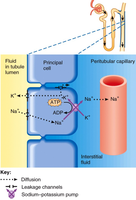
The collecting duct is the final site for reabsorption and secretion, involving Na+ leakage channels, principal cells, and intercalated cells.
Principal cells: Regulate Na+ and water reabsorption.
Intercalated cells: Regulate acid-base balance.
Hormonal Regulation of Kidney Function
RAA System, ADH, ANP & BNP
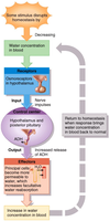
Hormones play a critical role in regulating kidney function and urine concentration. The renin-angiotensin-aldosterone (RAA) system increases Na+ and water reabsorption, antidiuretic hormone (ADH) increases water reabsorption via aquaporin-2 channels, and natriuretic peptides (ANP & BNP) decrease Na+ and water reabsorption.
RAA system: Increases blood volume and pressure.
ADH: Promotes water reabsorption in collecting ducts.
ANP & BNP: Promote excretion of Na+ and water.
Urine Concentration
Dilute vs. Concentrated Urine
The kidneys can produce dilute or concentrated urine depending on the body's needs. Concentrated urine formation involves an osmotic gradient, permeability changes, and the countercurrent multiplier mechanism.
Osmotic gradient: Created by Na+, Cl-, and urea.
Permeability: Regulated by ADH.
Countercurrent multiplier: Enhances concentration of urine.
Fluid, Electrolyte, and Acid-Base Balance
Fluid Compartments & Balance
Body fluids are distributed between extracellular fluid (ECF) and intracellular fluid (ICF). Fluid balance, electrolyte balance, and acid-base balance are essential for homeostasis.
ECF: Includes plasma and interstitial fluid.
ICF: Fluid within cells.
Fluid shift: Movement between compartments.
Regulation of Water/Solute Loss
Homeostasis monitors ECF losses and gains. Receptors monitor fluid and electrolytes indirectly. Active transport cannot move water directly.
NaCl loss: Can lead to hyponatremia or hypernatremia.
Hormonal control: Angiotensin II, aldosterone, ADH, natriuretic peptides.
Acid-Base Balance
Acids, Bases, and pH
Acids increase the concentration of hydrogen ions ([H+]), while bases decrease it. The body maintains pH within a narrow range (7.35–7.45) using buffer systems and compensation mechanisms.
Strong acids/bases: Completely dissociate in solution.
Weak acids/bases: Partially dissociate.
Buffer systems: Protein, phosphate, carbonic acid-bicarbonate.
Compensation: Respiratory and renal mechanisms.
Buffer Systems
Buffer systems help maintain pH by neutralizing excess acids or bases.
Protein buffer system: Uses amino acids to buffer changes in pH.
Phosphate buffer system: Buffers pH in intracellular fluid and urine.
Carbonic acid-bicarbonate buffer system: Important in plasma; involves the equation:
Acid-Base Disorders
Acid-base disorders include acidemia (low pH) and alkalemia (high pH). Compensation can be complete or partial, and disorders are classified as respiratory or metabolic.
Respiratory acidosis: Due to hypercapnia (high CO2).
Respiratory alkalosis: Due to hypocapnia (low CO2).
Metabolic acidosis: Causes include lactic acidosis, ketoacidosis, kidney damage, bicarbonate loss.
Metabolic alkalosis: Causes include alkaline tide.
Example: In diabetic ketoacidosis, excess ketone bodies lower blood pH, leading to metabolic acidosis.
Additional info: The notes have been expanded to provide academic context and explanations for each topic, including definitions, examples, and relevant equations.
