 뒤로
Urinary System: Structure and Function Study Guide
스터디 가이드 - 스마트 노트
 자료에 맞춘 맞춤형 노트, 핵심 정의, 예시, 맥락을 확장해 제공합니다.
Urinary System Overview
Functions of the Kidneys
The kidneys are essential organs responsible for maintaining homeostasis by regulating the composition and volume of body fluids. They perform several critical functions:
Regulation of water volume and solute concentration: The kidneys control the total amount of water and dissolved substances in the body.
Regulation of extracellular fluid (ECF) ion concentrations: They maintain proper levels of ions such as sodium, potassium, and calcium.
Acid-base balance: The kidneys ensure long-term regulation of blood pH.
Removal of metabolic wastes: They excrete waste products, toxins, and drugs from the body.
Endocrine functions: The kidneys produce hormones such as renin (regulates blood pressure), erythropoietin (stimulates red blood cell production), and activate vitamin D.
Gluconeogenesis: During prolonged fasting, the kidneys can produce glucose from non-carbohydrate sources.
Organs of the Urinary System
The urinary system consists of four main organs, each with a specific role in urine formation, transport, storage, and elimination:
Kidneys: Major excretory organs that filter blood and produce urine.
Ureters: Tubes that transport urine from the kidneys to the urinary bladder.
Urinary bladder: A muscular sac that temporarily stores urine.
Urethra: A tube that carries urine from the bladder to the outside of the body.
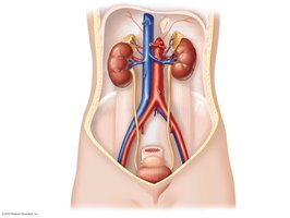
Anatomy of the Kidneys
Position and Structure
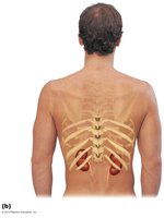
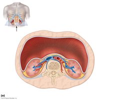
The kidneys are located against the posterior body wall, protected by the lower ribs and surrounded by supportive tissue layers. They are retroperitoneal, meaning they lie behind the peritoneal cavity.
Renal fascia: Outermost layer anchoring the kidney.
Perirenal fat capsule: Cushions and protects the kidney.
Fibrous capsule: Directly surrounds the kidney, providing protection.
Internal Anatomy
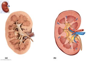
Each kidney has a complex internal structure that facilitates its function:
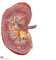
Renal cortex: The outer region where filtration occurs.
Renal medulla: The inner region containing renal pyramids.
Renal pelvis: Funnel-shaped structure that collects urine and channels it to the ureter.
Renal hilum: Entry/exit site for blood vessels, nerves, and ureter.
Major and minor calyces: Collect urine from the pyramids.
Blood and Nerve Supply
The kidneys receive a rich blood supply to filter and adjust blood composition:
Renal arteries: Deliver about 25% of cardiac output to the kidneys each minute.
Renal veins: Return filtered blood to the circulation.
Nerve supply: Provided by sympathetic fibers from the renal plexus.
Microscopic Anatomy: Nephrons
Structure and Function
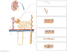
Nephrons are the structural and functional units of the kidney, responsible for urine formation. Each kidney contains over one million nephrons, each consisting of:
Renal corpuscle: Includes the glomerulus and Bowman's capsule, where filtration occurs.
Renal tubule: Processes filtrate into urine through reabsorption and secretion.
Classes of Nephrons
There are two main types of nephrons:
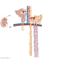
Cortical nephrons: Make up 85% of nephrons; located mostly in the cortex.
Juxtamedullary nephrons: Have long loops that extend deep into the medulla; crucial for producing concentrated urine.
Nephron Capillary Beds
Nephrons are associated with specialized capillary beds:
Glomerulus: Specialized for filtration; fed and drained by arterioles.
Peritubular capillaries: Adapted for absorption; surround renal tubules in the cortex.
Vasa recta: Long vessels parallel to nephron loops in juxtamedullary nephrons; important for urine concentration.
Juxtaglomerular Complex (JGC)
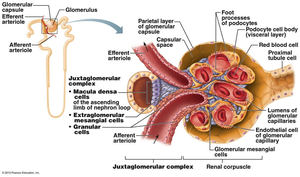
The JGC is a specialized structure involved in regulating filtrate formation and blood pressure. It consists of:
Macula densa: Chemoreceptors sensing NaCl content.
Granular cells: Mechanoreceptors that secrete renin.
Extraglomerular mesangial cells: Pass signals between macula densa and granular cells.
Urine Formation and Characteristics
Physical Characteristics
Urine is typically clear and ranges from pale to deep yellow due to the pigment urochrome. Its odor, pH, and specific gravity can vary:
Odor: Slightly aromatic when fresh; develops ammonia odor upon standing.
pH: Slightly acidic (~6), but can range from 4.5 to 8.0 depending on diet and health.
Specific gravity: 1.001 to 1.035, reflecting solute concentration.
Chemical Composition
Urine is composed of 95% water and 5% solutes, including:
Urea: From amino acid breakdown; largest solute component.
Uric acid: From nucleic acid metabolism.
Creatinine: From creatine phosphate metabolism.
Other solutes: Na+, K+, PO43–, SO42–, Ca2+, Mg2+, HCO3–.
Abnormal components (e.g., blood, proteins, WBCs, bile pigments) may indicate pathology.
Urine Transport, Storage, and Elimination
Ureters
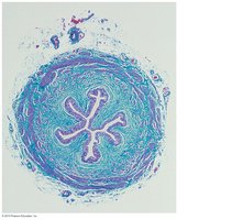
Ureters are muscular tubes that convey urine from the kidneys to the bladder. They begin at the renal pelvis and enter the bladder through the posterior wall. As bladder pressure increases, the distal ends of the ureters close to prevent backflow.
Urinary Bladder
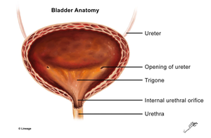
The urinary bladder is a muscular sac for temporary urine storage. It is retroperitoneal and located on the pelvic floor. The trigone is a smooth triangular area outlined by the openings for the ureters and urethra, where infections tend to persist.
Urethra
The urethra is a muscular tube that drains urine from the bladder. Its lining epithelium varies:
Transitional epithelium: Near the bladder.
Pseudostratified columnar epithelium: Most of the length.
Stratified squamous epithelium: Near the external urethral orifice.
There are two sphincters:
Internal urethral sphincter: Involuntary, smooth muscle at the bladder-urethra junction.
External urethral sphincter: Voluntary, skeletal muscle surrounding the urethra as it passes through the pelvic floor.
Homeostatic Imbalances
Kidney Infections
Infections can affect different parts of the kidney:
Pyelitis: Infection of the renal pelvis and calyces.
Pyelonephritis: Infection/inflammation of the entire kidney; usually treatable with antibiotics.
Summary Table: Urinary System Components and Functions
Component | Main Function |
|---|---|
Kidney | Filtration, regulation, hormone production |
Ureter | Transport urine to bladder |
Urinary Bladder | Temporary storage of urine |
Urethra | Elimination of urine from body |
