 뒤로
Urinary System: Tubular Reabsorption, Secretion, and Clinical Aspects
스터디 가이드 - 스마트 노트
 자료에 맞춘 맞춤형 노트, 핵심 정의, 예시, 맥락을 확장해 제공합니다.
The Urinary System
Overview
The urinary system is essential for maintaining homeostasis by regulating the composition and volume of blood, removing waste products, and controlling water and electrolyte balance. This section focuses on the processes of tubular reabsorption, secretion, urine concentration, and clinical evaluation of kidney function.
Tubular Reabsorption
Definition and Importance
Tubular reabsorption is the process by which the kidneys reclaim most of the filtrate from the nephron and return it to the bloodstream. This selective process ensures that essential nutrients and water are conserved, while waste products are excreted.
Selective Reabsorption: Almost all organic nutrients are reabsorbed; water and ion reabsorption are hormonally regulated.
Active and Passive Mechanisms: Includes both active (requiring energy) and passive (diffusion/osmosis) transport.
Sodium Reabsorption
Sodium (Na+) is the most abundant cation in the filtrate and is reabsorbed primarily by active transport.
Na+-K+ ATPase: Pumps Na+ out of the tubule cell into the interstitial space, facilitating its movement into peritubular capillaries.
Bulk Flow: Na+ is swept into capillaries by bulk flow.
Reabsorption of Nutrients, Water, and Ions
Water: Reabsorbed by osmosis, aided by aquaporins (water-filled pores).
Solutes: As water is reabsorbed, solute concentration increases, creating gradients for passive reabsorption of solutes such as fat-soluble substances, ions, and urea.
Lipid-soluble Drugs: These may be reabsorbed even if not desirable due to their solubility.
Transport Maximum (Tm)
Definition: The maximum rate at which a substance can be reabsorbed, determined by the number of available transport carriers.
Clinical Example: In hyperglycemia, glucose exceeds Tm and spills into urine (glycosuria).
Reabsorptive Capabilities of Renal Tubules and Collecting Ducts
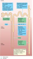
Proximal Convoluted Tubule (PCT): Site of most reabsorption; all nutrients, 65% of Na+ and water, many ions, almost all uric acid, and about half of urea.
Nephron Loop: Descending limb allows water to leave; ascending limb allows solutes to leave.
Distal Convoluted Tubule (DCT) and Collecting Duct: Reabsorption is hormonally regulated, especially by antidiuretic hormone (ADH), which increases water reabsorption by inserting aquaporins.
Tubular Secretion
Definition and Function
Tubular secretion is the process by which selected substances are moved from the blood into the filtrate for excretion. It is essentially reabsorption in reverse.
Substances Secreted: K+, H+, NH4+, creatinine, organic acids and bases.
Purpose: Helps control blood pH and eliminate substances not already in the filtrate.
Regulation of Urine Concentration and Volume
Countercurrent Mechanism
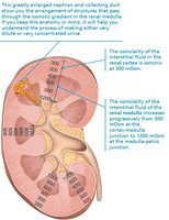
The kidneys regulate urine concentration and volume using a countercurrent mechanism, which involves fluid flowing in opposite directions in adjacent segments of the nephron loop.
Dehydration: Produces concentrated urine.
Overhydration: Produces dilute urine.
Countercurrent Exchange
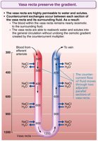
Vasa Recta: Blood vessels that preserve the osmotic gradient in the medulla by exchanging water and solutes.
Purpose: Allows the kidneys to produce urine of varying concentration.
Clinical Evaluation of Kidney Function
Urinalysis and Renal Clearance
Urinalysis: Examines urine for signs of disease or illegal substances.
Renal Clearance: Measures the volume of plasma cleared of a substance per unit time; used to determine glomerular filtration rate (GFR).
Clinical Homeostatic Imbalances
Chronic Renal Disease: GFR < 60 ml/min for 3 months; leads to accumulation of nitrogenous wastes and acidic blood.
Renal Failure: GFR < 15 ml/min; causes uremia, metabolic abnormalities, and requires dialysis or transplant.
Urine Composition and Characteristics
Chemical Composition
Water: 95% of urine.
Solutes: 5%, including urea, uric acid, creatinine, Na+, K+, PO43–, SO42–, Ca2+, Mg2+, HCO3–.
Abnormal Constituents: Blood proteins, WBCs, bile pigments may indicate pathology.
Abnormal Urinary Constituents
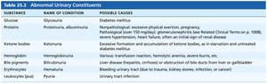
Glucose: Glycosuria; diabetes mellitus.
Proteins: Proteinuria; physical exertion, pregnancy, hypertension, heart failure, renal disease.
Ketone Bodies: Ketonuria; starvation, diabetes mellitus.
Hemoglobin: Hemoglobinuria; transfusion reaction, burns, hemolytic anemia.
Bile Pigments: Bilirubinuria; liver disease.
Leukocytes: Pyuria; urinary tract infection.
Substance | Name of Condition | Possible Causes |
|---|---|---|
Glucose | Glycosuria | Diabetes mellitus |
Proteins | Proteinuria, albuminuria | Excessive exertion, pregnancy, hypertension, heart failure, renal disease |
Ketone bodies | Ketonuria | Starvation, diabetes mellitus |
Hemoglobin | Hemoglobinuria | Transfusion reaction, hemolytic anemia, burns |
Bile pigments | Bilirubinuria | Liver disease |
Leukocytes (pus) | Pyuria | Urinary tract infection |
Physical Characteristics
Color: Pale to deep yellow due to urochrome; abnormal colors may indicate disease or diet.
Odor: Slightly aromatic; ammonia odor develops upon standing; altered by drugs or disease.
pH: Slightly acidic (~6); varies with diet and disease.
Specific Gravity: Ranges from 1.001 to 1.035.
Transport, Storage, and Elimination of Urine
Ureters
Ureters are slender tubes that convey urine from the kidneys to the bladder.
Structure: Three layers: mucosa (transitional epithelium), muscularis (smooth muscle), adventitia (connective tissue).
Function: Peristaltic waves propel urine; prevents backflow.
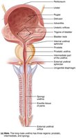
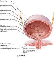
Urinary Bladder
The urinary bladder is a muscular sac for temporary storage of urine.
Location: On pelvic floor, posterior to pubic symphysis.
Capacity: Moderately full bladder holds ~500 ml; can hold up to twice that amount.
Structure: Contains rugae, expands during filling.
Pyelogram
A pyelogram is an imaging technique used to visualize the kidneys, ureters, and bladder. 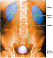
Urethra
The urethra is a muscular tube that drains the urinary bladder.
Internal Urethral Sphincter: Involuntary, smooth muscle.
External Urethral Sphincter: Voluntary, skeletal muscle.
Male Urethra: Carries both semen and urine; divided into prostatic, membranous, and spongy regions.
Clinical Homeostatic Imbalances
Urinary Tract Infections (UTIs)
Causes: Improper toilet habits, short female urethra, E. coli bacteria.
Symptoms: Dysuria, urgency, frequency, fever, cloudy or blood-tinged urine.
Treatment: Antibiotics.
Urinary Incontinence and Retention
Incontinence: Caused by weakened pelvic muscles; stress or overflow types.
Retention: Inability to expel urine; may require catheterization.
Congenital Abnormalities
Horseshoe Kidney: Fusion of kidneys; usually asymptomatic.
Polycystic Kidney Disease: Fluid-filled cysts interfere with function.
Developmental Aspects of the Urinary System
Infants and Elderly
Infants: Frequent micturition due to small bladders; incontinence normal until nervous system matures.
Elderly: Kidneys shrink, nephrons decrease; GFR reduced; loss of bladder tone causes nocturia and incontinence.
Key Equations
Renal Clearance Formula
Renal Clearance (C):
Where:
U = concentration of substance in urine
V = flow rate of urine formation
P = concentration of substance in plasma
Summary Table: Abnormal Urinary Constituents
Substance | Name of Condition | Possible Causes |
|---|---|---|
Glucose | Glycosuria | Diabetes mellitus |
Proteins | Proteinuria, albuminuria | Excessive exertion, pregnancy, hypertension, heart failure, renal disease |
Ketone bodies | Ketonuria | Starvation, diabetes mellitus |
Hemoglobin | Hemoglobinuria | Transfusion reaction, hemolytic anemia, burns |
Bile pigments | Bilirubinuria | Liver disease |
Leukocytes (pus) | Pyuria | Urinary tract infection |
