 뒤로
뒤로Amino Acids and Protein Structure: Properties, Organization, and Stabilizing Forces
스터디 가이드 - 스마트 노트
 자료에 맞춘 맞춤형 노트, 핵심 정의, 예시, 맥락을 확장해 제공합니다.
Amino Acids: Properties and Classification
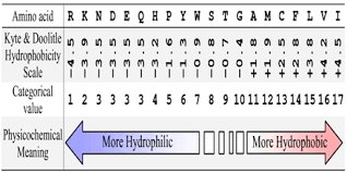
Hydrophobicity and Physicochemical Properties
Amino acids are the fundamental building blocks of proteins, each possessing unique side chains that determine their chemical behavior and role in protein structure. Hydrophobicity is a key property influencing protein folding and stability.
Hydrophobicity Scale: The Kyte & Doolittle scale ranks amino acids from most hydrophilic to most hydrophobic, affecting their location in protein structures (surface vs. core).
Physicochemical Meaning: Hydrophilic amino acids interact well with water, while hydrophobic amino acids prefer nonpolar environments.
Classification of Amino Acids
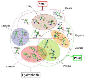
Amino acids can be grouped based on their side chain properties, which influence their function and placement in proteins.
Aliphatic: Non-aromatic, hydrophobic side chains (e.g., Alanine, Valine, Leucine, Isoleucine).
Aromatic: Contains a ring structure (e.g., Phenylalanine, Tyrosine, Tryptophan).
Polar: Side chains capable of hydrogen bonding (e.g., Serine, Threonine, Asparagine, Glutamine).
Charged: Acidic (Aspartate, Glutamate) or basic (Lysine, Arginine, Histidine) side chains.
Sulfur-containing: Cysteine and Methionine.
Small: Glycine, Alanine, and Proline (unique cyclic structure).
Examples and Applications
Cyclic: Proline (unique ring structure).
Aromatic: Phenylalanine, Tyrosine, Tryptophan.
Sometimes charged at physiological pH: Histidine.
Technically not hydrophobic: Glycine.
Polar or charged: Serine, Threonine, Aspartate, Glutamate, Lysine, Arginine.
Basic: Lysine, Arginine, Histidine.
Acidic: Aspartate, Glutamate.
Sulfur-containing: Cysteine, Methionine.
Aliphatic: Alanine, Valine, Leucine, Isoleucine.
Hydrophobic: Valine, Leucine, Isoleucine, Phenylalanine.
Aromatic hydrophobic and polar: Tyrosine (aromatic, polar hydroxyl group).
Hydrophobic polar and small: Glycine (small, not strongly hydrophobic).
Polypeptide Structure and Flexibility
Peptide Chain Structure and Limited Flexibility
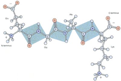
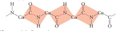
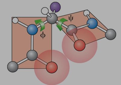
The peptide backbone consists of repeating units, with flexibility primarily at the alpha carbon (Cα) pivot points.
Peptide Bond: Formed between the carboxyl group of one amino acid and the amino group of another, resulting in a planar, rigid structure.
Flexibility: Rotation is allowed around the bonds adjacent to the Cα, but not around the peptide bond itself.
Stabilizing Forces in Protein Structure
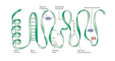
Protein structure is stabilized by several types of intermolecular forces:
Disulfide Bonds: Covalent bonds between cysteine residues, providing strong stabilization.
Hydrogen Bonds: Formed between backbone atoms or side chains, crucial for secondary structure.
Hydrophobic Effect: Drives nonpolar residues to the protein interior, stabilizing tertiary structure.
Van der Waals Forces: Weak interactions between closely packed atoms.
Electrostatic Interactions: Includes charge-charge, charge-dipole, and dipole-dipole interactions.
Secondary Structure: α-Helix
α-Helix Structure and Hydrogen Bonding
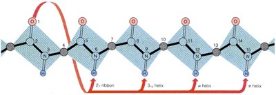
The α-helix is a common secondary structure stabilized by hydrogen bonds.
Hydrogen Bonding Pattern: Bonds form between the carbonyl oxygen of residue n and the amide hydrogen of residue n+4.
Residues per Turn: 3.6 residues per turn, with a rise of ~5.4 Å per turn.
Orientation: Carbonyls align along the helical axis; N-terminus is positively charged, C-terminus is negatively charged.
Side Chains: R-groups point outward from the helix, allowing interaction with the environment.
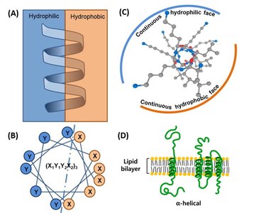
Amphipathic Helices
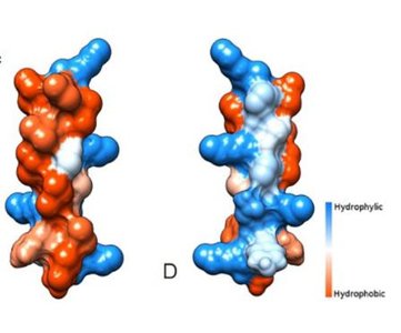
Some α-helices have distinct hydrophobic and hydrophilic faces, which is important for membrane proteins and antimicrobial peptides.
Amphipathic Helix: Nonpolar amino acids cluster on one side, polar/charged on the other.
Example: Pandinin 2 peptide forms an amphipathic helix, enabling it to interact with both bacterial membranes and red blood cells.
Secondary Structure: β-Sheet
β-Sheet Structure and Hydrogen Bonding
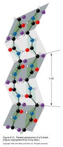
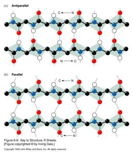
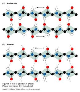
β-sheets are formed by extended polypeptide chains connected by hydrogen bonds between backbone atoms.
Antiparallel vs. Parallel: Strands can run in opposite (antiparallel) or same (parallel) directions.
Hydrogen Bonding: Bonds form between carbonyl oxygen and amide hydrogen of adjacent strands.
Characteristic Distance: Sheets are typically separated by ~7 Å.
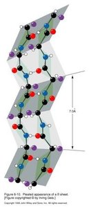
Comparison of α-Helix and β-Sheet Hydrogen Bonding
α-Helix: Hydrogen bonds are intrachain, connecting n to n+4 residues, oriented parallel to the helix axis.
β-Sheet: Hydrogen bonds are interchain, connecting adjacent strands, oriented perpendicular to the strand direction.
Protein Structure: Levels of Organization
Hierarchical Organization of Protein Structure
Protein structure is organized into four levels:
Primary: Amino acid sequence.
Secondary: Local structures (α-helix, β-sheet, turns).
Tertiary: Overall 3D folding of a single polypeptide.
Quaternary: Assembly of multiple polypeptide chains.
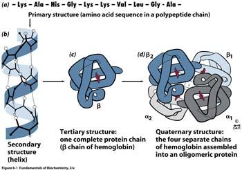
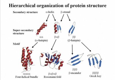
Supersecondary Structure and Motifs
Supersecondary structures are combinations of secondary elements, forming motifs such as β-hairpins, Greek key, and Rossmann fold.
Motifs: Simple structural patterns repeated in proteins.
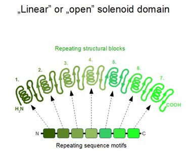
Domains: Independently folding units within a protein, often associated with specific functions.
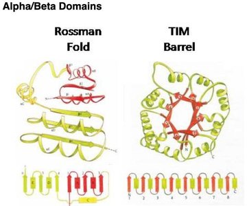
Alpha/Beta Domains: Rossmann Fold and TIM Barrel
Rossmann Fold: Common in nucleotide-binding proteins.
TIM Barrel: Found in many enzymes, active site at C-terminal end of β-strands.
Virus Assembly and Protein Structure
Virus capsids are assembled from protein subunits, demonstrating the importance of quaternary structure and intermolecular interactions.
Assembly: Involves nucleation, growth, and conformational changes.
Capsid: Complete viral shell formed by protein subunits.
Summary Table: Amino Acid Properties
Property | Example Amino Acids |
|---|---|
Cyclic | Proline |
Aromatic | Phenylalanine, Tyrosine, Tryptophan |
Sometimes charged at physiological pH | Histidine |
Technically not hydrophobic | Glycine |
Polar or charged | Serine, Threonine, Aspartate, Glutamate, Lysine, Arginine |
Basic | Lysine, Arginine, Histidine |
Acidic | Aspartate, Glutamate |
Sulfur-containing | Cysteine, Methionine |
Aliphatic | Alanine, Valine, Leucine, Isoleucine |
Hydrophobic | Valine, Leucine, Isoleucine, Phenylalanine |
Aromatic hydrophobic and polar | Tyrosine |
Hydrophobic polar and small | Glycine |
Key Equations
Peptide Bond Formation:
α-Helix Hydrogen Bond:
β-Sheet Hydrogen Bond: (i and j are on adjacent strands)
Additional info:
Histones are rich in basic amino acids (Lysine, Arginine) due to their role in binding negatively charged DNA via electrostatic interactions.
Arginine is more likely to be buried than Lysine due to its ability to form multiple hydrogen bonds and its less flexible side chain.
Protein denaturation by pH changes disrupts hydrogen bonds, electrostatic interactions, and sometimes disulfide bonds.
Site-directed mutagenesis of Lys in PEP carboxylase: Substitution with Arg (similar charge and size) would have the least effect; Glu (opposite charge) would have the greatest effect.
