 뒤로
뒤로Comprehensive Study Notes: Protein Structure and Function (Biochemistry)
스터디 가이드 - 스마트 노트
 자료에 맞춘 맞춤형 노트, 핵심 정의, 예시, 맥락을 확장해 제공합니다.
Proteins: Structure and Function
Peptide Bond Formation
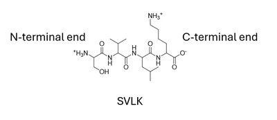
The peptide bond is a fundamental amide linkage formed by the condensation of two amino acids, resulting in the release of water. This bond is central to the formation of peptides and proteins, which can range from a few to thousands of amino acids in length. The average residue mass in a protein is about 110 Da, and the sequence of amino acids (primary structure) is always written from the N-terminal (amino group) to the C-terminal (carboxyl group).
Peptide vs. Protein: A peptide is generally a chain of fewer than 20 amino acids, while a protein or polypeptide is longer. This distinction is not rigid.
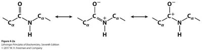
Partial Double Bond Character: The peptide bond exhibits resonance, giving it partial double bond character and restricting rotation around the C-N bond.
Protease Inhibitors: Drugs targeting proteases (e.g., HIV protease inhibitors) exploit the requirement for enzymatic hydrolysis of peptide bonds.
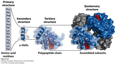
Levels of Protein Structure
Proteins exhibit hierarchical organization, with four distinct levels of structure that determine their function and interactions.
Primary Structure: The linear sequence of amino acids in a polypeptide chain.
Secondary Structure: Local spatial arrangements of the backbone, such as α-helices and β-sheets, stabilized by hydrogen bonds.
Tertiary Structure: The overall three-dimensional arrangement of a single polypeptide chain, stabilized by various weak interactions and sometimes disulfide bonds.
Quaternary Structure: The assembly of multiple polypeptide chains (subunits) into a functional protein complex.
Primary Structure
Peptide Bond Resonance and Rotation
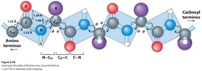
The peptide bond's resonance restricts rotation around the C-N bond, while rotation is permitted around the bonds to the α-carbon (phi and psi angles). These angles are critical in determining the possible conformations of the polypeptide backbone.
Phi (φ) Angle: Rotation around the N–Cα bond.
Psi (ψ) Angle: Rotation around the Cα–C bond.
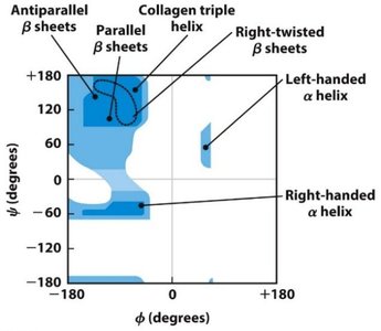
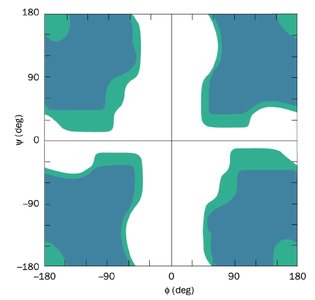
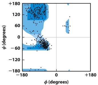
Ramachandran Plot: Visualizes the allowed regions of φ and ψ angles for amino acids in proteins, highlighting steric constraints.
Ramachandran Plots
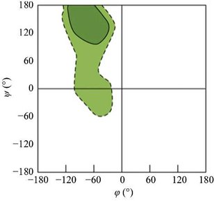
Ramachandran plots show the sterically allowed regions for backbone dihedral angles in proteins. Glycine, due to its small side chain, is less restricted, while proline is more restricted due to its cyclic structure.
Secondary Structure
α-Helix
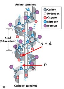
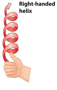
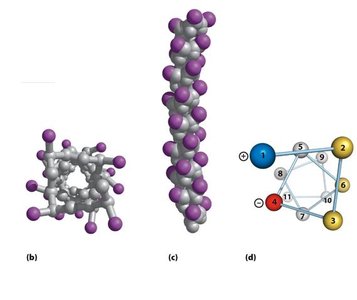
The α-helix is a right-handed helical structure stabilized by hydrogen bonds between the backbone NH group of residue n and the carbonyl oxygen of residue n+4. There are approximately 3.6 residues per turn, and side chains project outward from the helix axis.
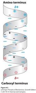
Helical Dipole: The α-helix has a macroscopic dipole, with the amino end more positive and the carboxyl end more negative.
Helical Wheel: Used to visualize the spatial arrangement of side chains, often revealing amphipathic helices with hydrophobic and hydrophilic faces.
Residue Preferences: Glycine and tryptophan are rarely found in α-helices due to their unique properties.
β-Sheets
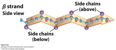
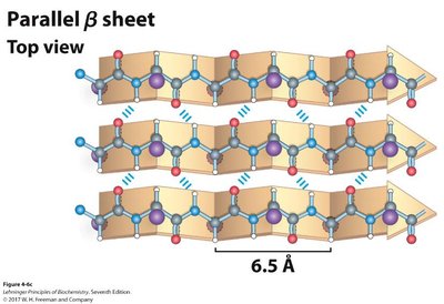
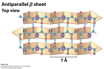
β-sheets are formed by hydrogen bonding between backbone amides and carbonyls of adjacent β-strands. Strands can be parallel (same N-to-C direction) or antiparallel (opposite directions), with antiparallel sheets having stronger, more linear hydrogen bonds.
R Group Orientation: Side chains alternate above and below the plane of the sheet.
Structural Role: β-sheets are common in structural proteins and contribute to protein stability.
β-Turns
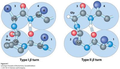
β-turns allow the polypeptide chain to reverse direction, often connecting strands of antiparallel β-sheets. They are stabilized by a hydrogen bond between the carbonyl oxygen of residue 1 and the amide proton of residue 4. Proline and glycine are commonly found in β-turns due to their unique conformational properties.
Proline Isomerization
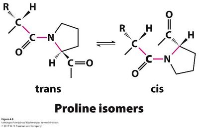
While most peptide bonds are in the trans configuration, about 6% of peptide bonds preceding proline are in the cis configuration. Proline isomerases catalyze the interconversion between these forms, which can be important for protein folding.
Tertiary Structure
Classification of Proteins
Proteins can be classified based on their overall structure and solubility:
Fibrous Proteins: Elongated, insoluble, and primarily structural (e.g., α-keratin, collagen, silk fibroin).
Globular Proteins: Compact, water-soluble, and functionally diverse (e.g., enzymes, myoglobin).
Intrinsically Disordered Proteins: Lack stable tertiary structure under physiological conditions but are functionally important.
Membrane Proteins: Associated with or embedded in cell membranes.
Peripheral Membrane Proteins: Associate with membrane surfaces via non-covalent interactions or lipidation.
Fibrous Proteins
Fibrous proteins provide structural support and are characterized by repetitive secondary structures. Examples include α-keratin (hair, nails), collagen (connective tissue), and silk fibroin (silk).
Structure | Characteristics | Examples of Occurrence |
|---|---|---|
α Helix, cross-linked by disulfide bonds | Tough, insoluble protective structures of varying hardness and flexibility | α-Keratin of hair, feathers, nails |
β Conformation | Soft, flexible filaments | Silk fibroin |
Collagen triple helix | High tensile strength, without stretch | Collagen of tendons, bone matrix |
α-Keratin
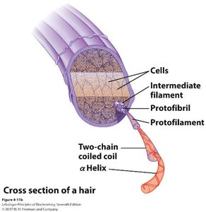
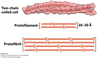
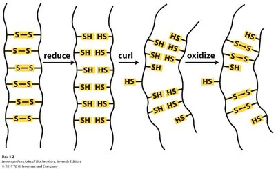
α-Keratin forms coiled-coil structures that assemble hierarchically into protofilaments, protofibrils, and intermediate filaments, providing mechanical strength to hair and other tissues. Disulfide bonds between helices contribute to the physical properties of hair (e.g., straight vs. curly).
Collagen
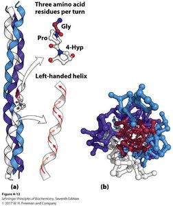
Collagen is a major component of connective tissue, consisting of three left-handed helical chains that form a right-handed triple helix. It is rich in glycine and proline, and contains hydroxyproline, a post-translationally modified residue essential for stability. Vitamin C is required for hydroxyproline formation; deficiency leads to scurvy.
Silk Fibroin
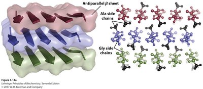
Silk fibroin is composed of antiparallel β-sheets with tightly packed glycine and alanine residues, resulting in a material with remarkable tensile strength and extensibility.
Globular Proteins
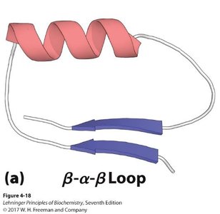
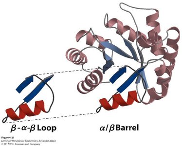
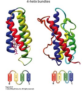
Globular proteins are water-soluble and exhibit complex tertiary structures formed by combinations of secondary structure motifs (e.g., β-α-β loops, α/β barrels, β-barrels, immunoglobulin folds). These motifs are critical for protein function and specificity.
Intrinsically Disordered Proteins
Some proteins or regions lack a fixed tertiary structure and are termed intrinsically disordered. These regions are often rich in lysine, arginine, glutamate, and proline, and can adopt different conformations depending on their binding partners. The tumor suppressor p53 is a classic example, with disordered N- and C-terminal regions that mediate diverse protein-protein interactions.
Quaternary Structure
Quaternary structure arises when multiple polypeptide chains (subunits) assemble into a functional protein complex. Subunits can be identical (homo-) or different (hetero-), and the arrangement allows for cooperative function, as seen in hemoglobin (a tetramer of four subunits).
Protein Stability and Folding
Folding and Denaturation
The native state of a protein is its functional, folded conformation, determined by its primary sequence. Denaturation is the loss of structural integrity and function, caused by heat, pH extremes, organic solvents, or chaotropic agents. Protein folding is driven by the hydrophobic effect, with hydrophobic residues collapsing inward, followed by formation of secondary and tertiary structures.
Proteostasis: The cellular processes that control protein synthesis, folding, and degradation.
Folding Energy Landscape
The folding process can be visualized as a funnel-shaped energy landscape, with many possible conformations at high energy and the native state at the lowest energy. Proteins may become trapped in intermediate states, requiring energy input (e.g., from chaperones) to reach the native state.
Chaperones and Protein Folding
Chaperone proteins assist in the proper folding of other proteins and prevent aggregation. The Hsp70/Hsp40 system binds misfolded proteins, preventing aggregation and allowing refolding. If unsuccessful, the GroEL/ES system provides an isolated environment for refolding, using ATP hydrolysis to drive conformational changes.
Summary Table: Secondary Structures and Properties of Some Fibrous Proteins
Structure | Characteristics | Examples of Occurrence |
|---|---|---|
α Helix, cross-linked by disulfide bonds | Tough, insoluble protective structures of varying hardness and flexibility | α-Keratin of hair, feathers, nails |
β Conformation | Soft, flexible filaments | Silk fibroin |
Collagen triple helix | High tensile strength, without stretch | Collagen of tendons, bone matrix |
Key Equations
Average Protein Molecular Weight:
Average mRNA Molecular Weight:
Clinical Connections
Vitamin C and Scurvy: Vitamin C is required for hydroxyproline formation in collagen. Deficiency leads to destabilized collagen and connective tissue breakdown (scurvy).
p53 and Cancer: Loss of one functional copy of the p53 gene (as in Li-Fraumeni syndrome) predisposes individuals to multiple tumors due to impaired cell cycle regulation.
Cystic Fibrosis: The ΔF508 mutation in CFTR leads to misfolding and degradation of the protein, not direct loss of function.
Summary
Understanding protein structure, from the primary sequence to quaternary assemblies, is essential for grasping protein function, stability, and the molecular basis of many diseases. The interplay of weak forces, post-translational modifications, and chaperone-mediated folding ensures that proteins achieve and maintain their functional conformations.
