 뒤로
뒤로DNA: The Genetic Material – Structure, Evidence, and Function
스터디 가이드 - 스마트 노트
 자료에 맞춘 맞춤형 노트, 핵심 정의, 예시, 맥락을 확장해 제공합니다.
DNA: The Genetic Material
Experimental Evidence for DNA as Genetic Material
The identification of DNA as the genetic material was established through a series of landmark experiments. These experiments demonstrated that DNA, not protein, carries genetic information in cells.
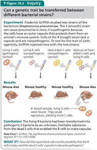
Griffith's Transformation Experiment (1928): Demonstrated that a "transforming principle" from dead pathogenic bacteria could genetically alter non-pathogenic bacteria, suggesting the transfer of genetic material.
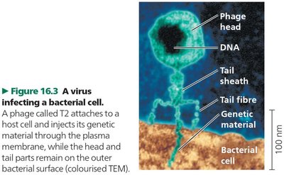
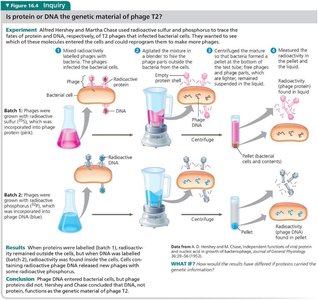
Hershey-Chase Experiment (1952): Used radioactive labeling to show that DNA, not protein, is injected by bacteriophages into bacteria, confirming DNA as the genetic material.
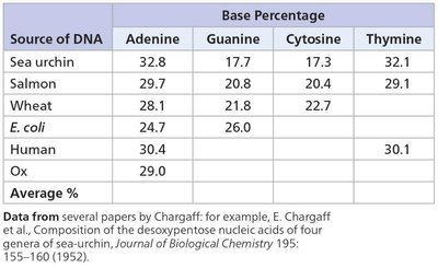
Chargaff's Rules: Established that DNA composition varies between species but maintains specific ratios: the amount of adenine equals thymine, and guanine equals cytosine.
Chargaff's Rules and DNA Composition
Chargaff's analysis of DNA from various organisms revealed consistent base pairing ratios, which were critical for understanding DNA structure.
Base Pairing: In DNA, the number of adenine (A) residues equals thymine (T), and guanine (G) equals cytosine (C).
Species Variation: The overall base composition varies between species, but the ratios remain constant within a species.
Source of DNA | Adenine (%) | Guanine (%) | Cytosine (%) | Thymine (%) |
|---|---|---|---|---|
Sea urchin | 32.8 | 17.7 | 17.3 | 32.1 |
Salmon | 29.7 | 20.8 | 20.4 | 29.1 |
Wheat | 28.1 | 21.8 | 22.7 | 27.4 |
E. coli | 24.7 | 26.0 | 25.7 | 23.6 |
Human | 30.4 | 19.9 | 19.8 | 30.1 |
Ox | 29.0 | 22.0 | 22.0 | 27.0 |
X-ray Diffraction and DNA Structure
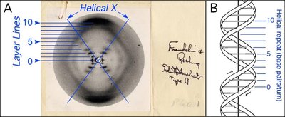
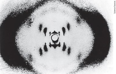
X-ray diffraction studies by Rosalind Franklin and Maurice Wilkins provided crucial evidence for the helical structure of DNA. The patterns revealed the double-stranded nature and periodicity of the helix.
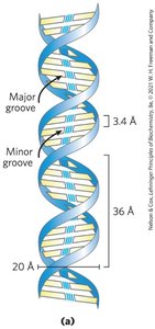
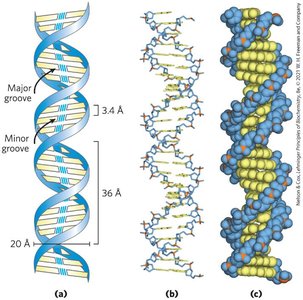
Helical Structure: DNA is a double helix with a width of about 20 Å and periodicities of 3.4 Å and 34 Å along its axis.
Sugar-Phosphate Backbone: Located on the outside, facing the aqueous environment.
Discovery of the DNA Double Helix
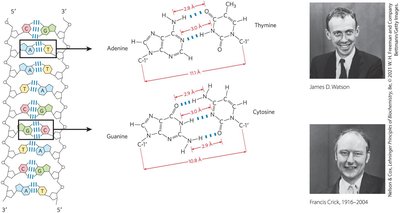
Watson and Crick proposed the three-dimensional structure of DNA in 1953, integrating X-ray diffraction data and Chargaff's rules. Their model explained how DNA could store and transmit genetic information.
Double Helix: Two antiparallel strands wound around a common axis.
Complementary Base Pairing: A pairs with T (two hydrogen bonds), G pairs with C (three hydrogen bonds).
Major and Minor Grooves: The helix has alternating grooves, important for protein-DNA interactions.
Nucleotides and Nucleic Acid Structure
Nucleotide Structure
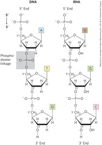
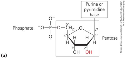
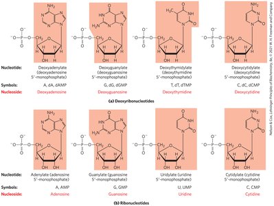
Nucleotides are the basic building blocks of nucleic acids. Each nucleotide consists of a pentose sugar, a nitrogenous base, and a phosphate group.
Pentose Sugar: Ribose in RNA (2' OH), deoxyribose in DNA (2' H).
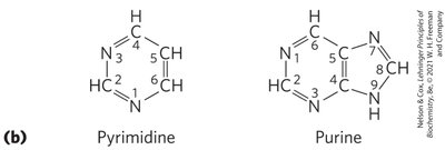
Nitrogenous Bases: Purines (A, G) and pyrimidines (C, T in DNA; C, U in RNA).
Nucleoside: Sugar + base (no phosphate).
Nucleotide: Sugar + base + phosphate.
Polynucleotide Chains and Backbone
Nucleotides are linked by phosphodiester bonds, forming a linear, directional polymer. The backbone is negatively charged and stable in DNA, less so in RNA.
Phosphodiester Linkage: Connects 5' phosphate to 3' hydroxyl of adjacent nucleotide.
Directionality: Sequence is read from 5' to 3'.
Linear Structure: No branching or cross-links.
DNA Double Helix: Structural Features
Watson-Crick Model
The double helix is stabilized by base-stacking interactions, hydrogen bonds, and metal cations. The two strands are antiparallel and complementary.
Base Stacking: Hydrophobic interactions between planar bases.
Hydrogen Bonds: Between complementary bases.
Metal Cations: Bind to the negatively charged backbone.
DNA Replication: Semi-Conservative Mechanism
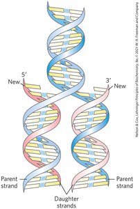
DNA replication involves strand separation, with each strand serving as a template for synthesis of a new complementary strand. This ensures the retention of the original sequence.
Semi-Conservative Replication: Each new DNA molecule contains one parent and one daughter strand.
DNA Polymerases: Catalyze the synthesis of new DNA strands.
DNA Function and Co-linearity
Co-linearity of DNA, mRNA, and Protein
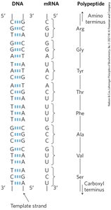
The nucleotide sequence of DNA is transcribed into mRNA, which is then translated into the amino acid sequence of a protein. This direct relationship is known as co-linearity.
Transcription: DNA coding sequence is copied into mRNA.
Translation: mRNA codons specify amino acids in proteins.
Protein Function: Determined by the order of amino acids.
Three-Dimensional Forms of DNA
B, A, and Z Forms of DNA
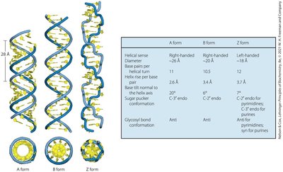
DNA can adopt several three-dimensional forms, depending on environmental conditions and sequence.
B Form: Most common, right-handed helix, 10.5 base pairs per turn.
A Form: Right-handed, wider helix, occurs in low water conditions.
Z Form: Left-handed, zig-zag backbone, occurs with alternating CG sequences.
Form | Helical Sense | Diameter (Å) | Base Pairs/Turn | Helix Rise/Pair (Å) |
|---|---|---|---|---|
A | Right-handed | 26 | 11 | 2.6 |
B | Right-handed | 20 | 10.5 | 3.4 |
Z | Left-handed | 18 | 12 | 3.7 |
Unusual Local Secondary Structures
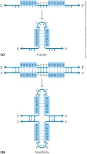
DNA and RNA can form local secondary structures such as palindromes, hairpins, and cruciforms, which are important for gene regulation and restriction enzyme sites.
Palindromes: Sequences that read the same forward and backward.
Hairpins: Formed by single-stranded nucleic acids folding back on themselves.
Cruciforms: Double-stranded structures formed by palindromic sequences.
Levels of DNA/RNA Structure
Primary, Secondary, and Tertiary Structure
Nucleic acids exhibit hierarchical structural organization:
Primary Structure: Covalent bonding and nucleotide sequence.
Secondary Structure: Helical forms, hairpins, cruciforms.
Tertiary Structure: Chromatin/chromosome folding, complex RNA folding.
Example: tRNA secondary and tertiary structures are critical for its function in translation.
Additional info: These notes are based on Lehninger Biochemistry (Nelson and Cox, Ed. VIII) and Campbell Biology (Ed. XII), focusing on Chapter 8 and Chapter 16, respectively.
