 뒤로
뒤로Enzyme Structure, Function, Kinetics, and Regulation: Comprehensive Study Notes
스터디 가이드 - 스마트 노트
 자료에 맞춘 맞춤형 노트, 핵심 정의, 예시, 맥락을 확장해 제공합니다.
Enzyme Structure and General Properties
Definition and Role of Enzymes
Enzymes are biological catalysts, primarily proteins, that accelerate chemical reactions without being consumed in the process. They act on specific substrates to produce products, often with remarkable specificity and efficiency.
- Catalyst: Increases reaction rate without being altered.
- Substrate: The molecule upon which an enzyme acts.
- Reaction specificity: Enzymes produce stereospecific products with minimal by-products.
- Regulation: Enzyme activity can be modulated by various mechanisms.
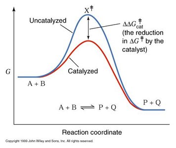
Energetics of Enzyme Catalysis
Enzymes lower the activation energy (ΔG‡) required for a reaction, thereby increasing the rate at which equilibrium is reached. However, they do not alter the overall change in Gibbs free energy (ΔG) or the equilibrium position.
- ΔG: Change in Gibbs free energy; determines spontaneity.
- ΔG‡: Activation energy; lowered by enzymes.
- Induced fit: Enzyme changes conformation to better bind substrate and stabilize the transition state.
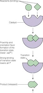
Enzyme Mechanisms and Active Sites
Active Site and Substrate Binding
The active site of an enzyme is a specialized region where substrate binding and catalysis occur. Folding of the protein brings together key side chains and backbone groups to form this site.
- Proximity and orientation: Substrate is positioned optimally for reaction.
- Transition state stabilization: Enzyme binds tightly to the transition state, lowering activation energy.
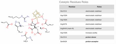
- Acid-base catalysis: Amino acid side chains (e.g., His, Cys, Tyr, Asp, Glu, Lys) donate or accept protons.
- Covalent catalysis: Temporary covalent bond forms between enzyme and substrate (e.g., Ser, Tyr, Cys, Lys, His).
- Cofactors: Non-protein molecules (e.g., Zn²⁺) assist in catalysis.
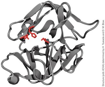
Specificity and Recognition
Enzyme specificity is determined by the chemical properties and geometry of the substrate binding pocket.
- Substrate recognition: Enzyme site matches substrate shape and charge.
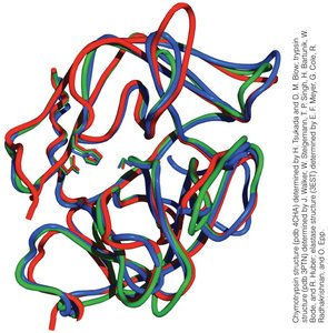
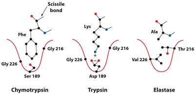
- Example: Chymotrypsin, trypsin, and elastase have similar structures but different substrate preferences due to their specificity pockets. 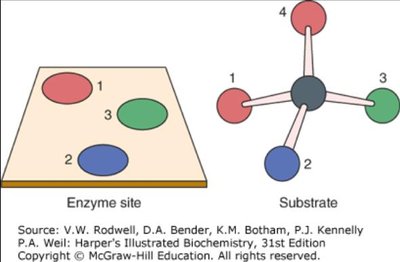
Chemical Strategies in Enzyme Catalysis
Nucleophilic and Electrophilic Catalysis
Enzymes utilize nucleophilic and electrophilic groups to facilitate bond formation and cleavage.
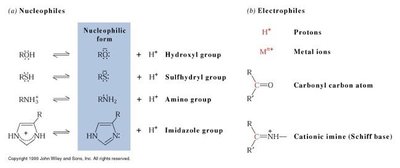
- Nucleophiles: Donate electron pairs (e.g., hydroxyl, sulfhydryl, amine, imidazole groups).
- Electrophiles: Accept electron pairs (e.g., protons, metal ions, carbonyl carbon, cationic imines).
Example: Aconitase Mechanism
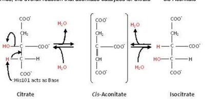
Aconitase isomerizes citrate to isocitrate via dehydration and rehydration steps, using a [4Fe–4S] cluster as a cofactor.
- Dehydration: Removes H from C2 and OH from C3 to form cis-aconitate.
- Rehydration: Adds water across the double bond, moving OH to C2 and H to C3.
- Net effect: Converts a tertiary alcohol to a secondary alcohol.
Condensation and Hydrolysis Reactions
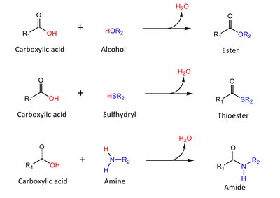
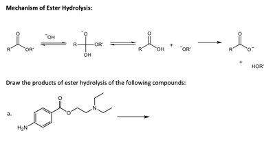
- Condensation: Two molecules combine, releasing water.
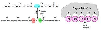
- Hydrolysis: Bond is broken by addition of water.
Proteolytic Enzymes: Serine Proteases
Chymotrypsin Mechanism and Specificity
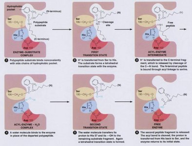
Chymotrypsin is a serine protease that catalyzes the hydrolysis of peptide bonds, especially after aromatic residues (Phe, Trp, Tyr).
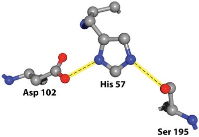
- Catalytic triad: Ser195, His57, Asp102 cooperate to activate Ser for nucleophilic attack.
- Specificity pocket: Determines substrate preference. 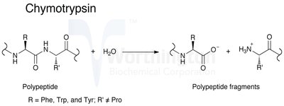
Enzyme Kinetics
Michaelis-Menten Model
The Michaelis-Menten equation describes the relationship between substrate concentration and reaction velocity for many enzymes. - Equation: - Km: Substrate concentration at which velocity is half-maximal; reflects enzyme affinity for substrate. - kcat: Turnover number; maximum number of substrate molecules converted per enzyme per second. - kcat/Km: Catalytic efficiency; higher values indicate more efficient enzymes. 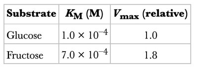
Substrate | KM (M) | Vmax (relative) |
|---|---|---|
Glucose | 1.0 × 10⁻⁴ | 1.0 |
Fructose | 7.0 × 10⁻⁴ | 1.8 |
Enzyme Inhibition
Competitive Inhibition
Competitive inhibitors bind to the active site, preventing substrate binding. - Effect: Apparent Km increases; Vmax remains unchanged. - Equation: 
Non-Competitive Inhibition
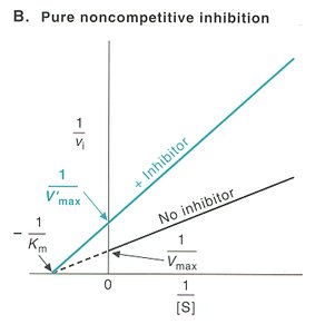
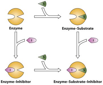
Non-competitive inhibitors bind to a site other than the active site, affecting enzyme activity regardless of substrate binding. - Effect: Vmax decreases; Km remains unchanged. 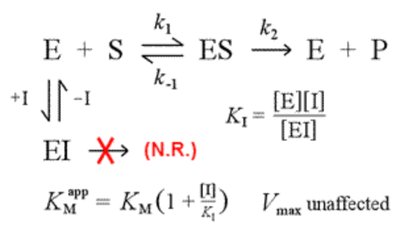
Uncompetitive Inhibition
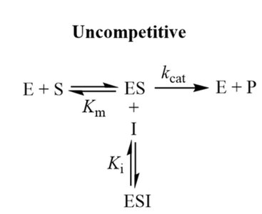
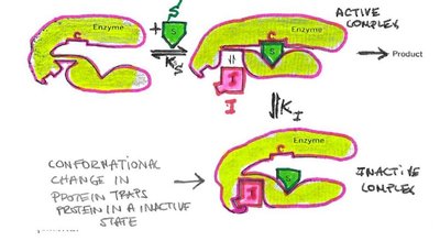
Uncompetitive inhibitors bind only to the enzyme-substrate complex, decreasing both Km and Vmax. 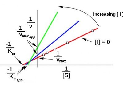
Mixed Inhibition
Mixed inhibitors bind to both the enzyme and the enzyme-substrate complex, affecting both Km and Vmax in variable ways. 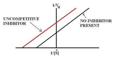
Non Michaelis-Menten Kinetics and Allostery
Allosteric Enzymes and Regulation
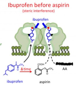
Allosteric enzymes do not follow Michaelis-Menten kinetics and are regulated by effectors that bind at sites other than the active site. - Allosteric inhibition: Effector decreases enzyme activity. - Allosteric activation: Effector increases enzyme activity. 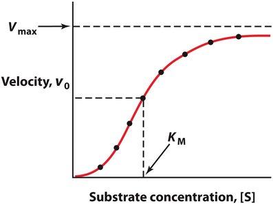
Metabolic Regulation of Enzymes
Mechanisms of Regulation
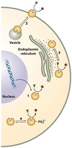
Enzyme activity is regulated at multiple levels, including synthesis, degradation, localization, allosteric regulation, covalent modification, and feedback inhibition. - Transcriptional regulation: Changes in enzyme gene expression. - Post-translational modification: Phosphorylation, acetylation, etc. - Feedback inhibition: End product inhibits pathway enzyme. 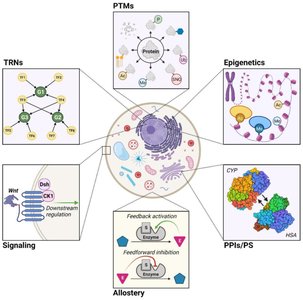
Summary Table: Types of Enzyme Inhibition
Type | Binding Site | Km (apparent) | Vmax (apparent) |
|---|---|---|---|
Competitive | Enzyme only | Increases | Unchanged |
Pure Noncompetitive | Enzyme and ES equally | Unchanged | Decreases |
Uncompetitive | ES only | Decreases | Decreases |
Mixed | Enzyme and ES unequally | Increases or decreases | Decreases |
Key Equations
Michaelis-Menten Equation
Competitive Inhibition
Lineweaver-Burk Plot
Conclusion
These notes provide a comprehensive overview of enzyme structure, function, kinetics, inhibition, and regulation, with relevant examples and diagrams to reinforce key concepts. Enzymes are central to biochemistry, and understanding their mechanisms and regulation is essential for advanced study and practical applications in medicine and biotechnology.
