 뒤로
뒤로Protein Binding, Molecular Recognition, and Allostery: Hemoglobin and Myoglobin
스터디 가이드 - 스마트 노트
 자료에 맞춘 맞춤형 노트, 핵심 정의, 예시, 맥락을 확장해 제공합니다.
Protein Binding, Molecular Recognition, and Allostery
Introduction
Protein binding and molecular recognition are fundamental concepts in biochemistry, illustrating how proteins interact with specific ligands to perform biological functions. Hemoglobin and myoglobin serve as classic examples, demonstrating specificity, cooperativity, and allosteric regulation in oxygen transport and storage.
Globins: Oxygen-Binding Proteins
Myoglobin
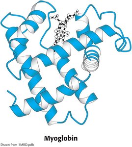
Myoglobin is a heme-containing monomeric protein found primarily in muscle tissue. Its main function is to store oxygen for use during periods of high metabolic demand.
Structure: Single polypeptide chain with a heme prosthetic group.
Function: Facilitates oxygen storage in muscle cells.
Hemoglobin
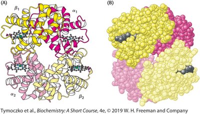
Hemoglobin is the major protein in red blood cells, responsible for transporting oxygen from the lungs to tissues throughout the body.
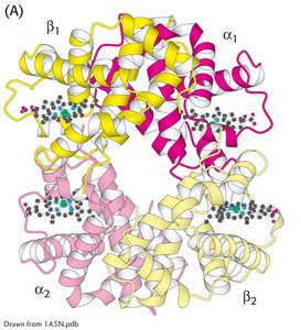
Structure: Tetramer composed of two α and two β subunits (α2β2).
Function: Oxygen transport in the bloodstream.
Heme: The Oxygen-Binding Prosthetic Group
Structure and Function of Heme
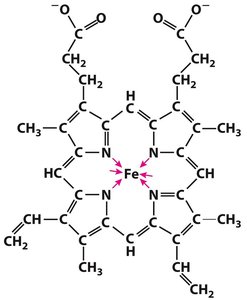
The heme group is a prosthetic group essential for oxygen binding in both myoglobin and hemoglobin. It consists of a protoporphyrin ring coordinated to a central iron ion (Fe2+).
Prosthetic Group: A non-polypeptide unit tightly bound to a protein, required for its biological activity.
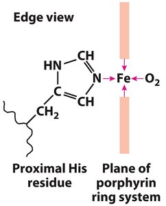
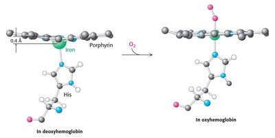
Iron Coordination: The iron ion forms six coordination bonds—four with nitrogen atoms in the porphyrin ring, one with a proximal histidine residue, and one with oxygen.
Oxygen Binding and Structural Changes
When oxygen binds to the heme iron, the iron ion moves into the plane of the porphyrin ring, inducing a conformational change in the protein. This movement is critical for the function of hemoglobin and myoglobin.
Protein Structure Comparison: Myoglobin vs. Hemoglobin
Primary and Tertiary Structure
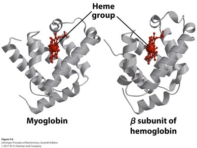
Despite differences in their primary sequences, myoglobin and the subunits of hemoglobin share remarkably similar tertiary structures, reflecting their common evolutionary origin and function.
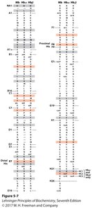
Conserved Residues: Key amino acids involved in heme binding and oxygen coordination are conserved across globins.
Oxygen Binding Properties
Binding Curves and Cooperativity
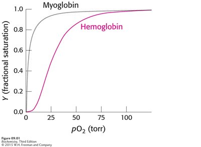
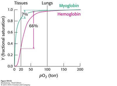
Myoglobin exhibits a hyperbolic oxygen-binding curve, indicating a simple, non-cooperative binding mechanism. In contrast, hemoglobin displays a sigmoidal curve, characteristic of cooperative binding—where the binding of one oxygen molecule increases the affinity for subsequent oxygen molecules.
Cooperativity: A form of allosteric regulation where ligand binding at one site affects binding at other sites.
Allosteric Regulation of Hemoglobin
Quaternary Structure and Conformational States
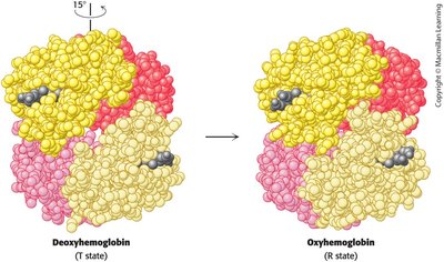
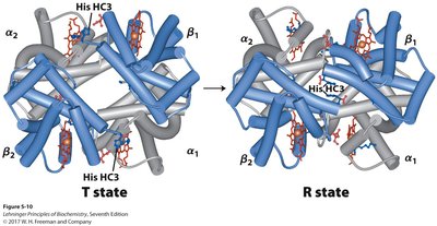
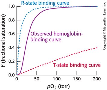
Hemoglobin exists in two major quaternary conformations: the T (tense) state with low oxygen affinity and the R (relaxed) state with high oxygen affinity. Oxygen binding induces a shift from the T to the R state, facilitating cooperative binding.
T State: Stabilized in the absence of oxygen; lower affinity for O2.
R State: Stabilized upon oxygen binding; higher affinity for O2.
Observed Binding Curve
The observed oxygen-binding curve for hemoglobin is a combination of the curves for the T and R states, resulting in a sigmoidal shape that reflects cooperative binding.
Allosteric Effectors of Hemoglobin
2,3-Bisphosphoglycerate (2,3-BPG)
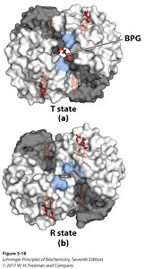
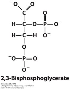
2,3-BPG is a key allosteric effector that binds to the central cavity of deoxyhemoglobin (T state), stabilizing it and reducing hemoglobin's affinity for oxygen. This facilitates oxygen release in tissues.
Binding Site: Central cavity between β subunits in the T state.
Physiological Role: Enhances oxygen delivery under conditions of low oxygen availability (e.g., high altitude, exercise).
Bohr Effect: pH and CO2 Regulation
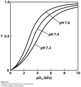
The Bohr effect describes how decreases in pH (increased H+ concentration) and increases in CO2 concentration reduce hemoglobin's affinity for oxygen, promoting oxygen release in metabolically active tissues.
Mechanism: H+ and CO2 bind to hemoglobin, stabilizing the T state and facilitating O2 release.
Equation:
Physiological Significance: Enhances oxygen delivery where it is most needed.
Summary Table: Comparison of Myoglobin and Hemoglobin
Property | Myoglobin | Hemoglobin |
|---|---|---|
Structure | Monomer | Tetramer (α2β2) |
Location | Muscle | Red blood cells |
Function | O2 storage | O2 transport |
O2 Binding Curve | Hyperbolic | Sigmoidal (cooperative) |
Allosteric Regulation | No | Yes (2,3-BPG, H+, CO2) |
