 뒤로
Protein Function: Myoglobin, Hemoglobin, and Antibodies
스터디 가이드 - 스마트 노트
 자료에 맞춘 맞춤형 노트, 핵심 정의, 예시, 맥락을 확장해 제공합니다.
Protein Function: Myoglobin and Hemoglobin
Myoglobin: Structure and Function
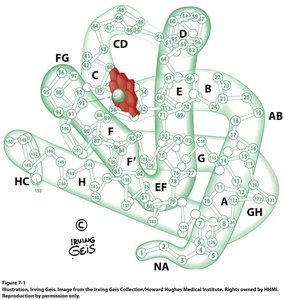
Myoglobin is a small, globular protein found in muscle tissue, primarily responsible for oxygen storage and transport within muscle cells. It consists of 153 amino acid residues arranged in eight α-helices, forming a compact tertiary structure.
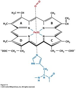
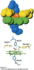
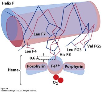
Oxygen Binding: Myoglobin contains a single heme prosthetic group, which is a porphyrin derivative with four pyrrole rings. The central iron (Fe2+) atom binds oxygen reversibly.
Prosthetic Group: The heme group is not covalently attached but is stabilized by noncovalent interactions with the protein.
Ligand Binding: Besides O2, molecules like CO, NO, and H2S can bind with higher affinity, which can be toxic.
Color Change: Oxygenation alters the electronic state of the heme, causing color changes (dark purple in veins, red in arteries).
Metmyoglobin: Oxidation of Fe2+ to Fe3+ forms metmyoglobin, responsible for the brown color of old meat.
Oxygen Binding Curve and Dissociation Constant
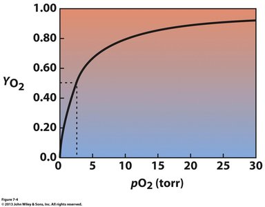
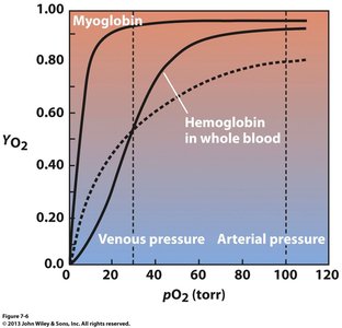
The binding of oxygen to myoglobin follows a hyperbolic curve, described by the dissociation constant (K) and fractional saturation (YO2):
Dissociation Constant (K):
Fractional Saturation (YO2):
p50: The partial pressure of O2 at which myoglobin is 50% saturated (p50 = 2.8 torr).
Hyperbolic Curve: Indicates non-cooperative binding typical of single-site proteins.
Hemoglobin: Structure and Function
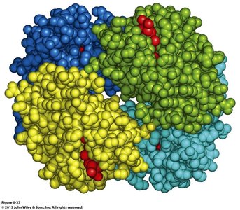
Hemoglobin is a tetrameric protein found in red blood cells, responsible for oxygen transport in the blood. It consists of two α and two β subunits, each containing a heme group.
Quaternary Structure: Hemoglobin forms a dimer of αβ protomers, allowing cooperative binding of oxygen.
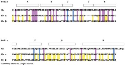
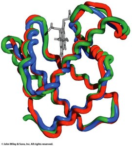
Sequence and Structure: Despite only ~18% sequence identity with myoglobin, hemoglobin's subunits have similar secondary and tertiary structures.
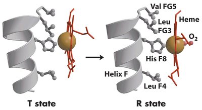
Oxygen Binding: The binding of oxygen induces a conformational change from the T (tense, deoxy) state to the R (relaxed, oxy) state, increasing affinity for O2.
Oxygen Binding Curve and Cooperativity
Hemoglobin exhibits a sigmoidal oxygen binding curve, indicative of cooperative binding. This allows efficient oxygen loading in the lungs and unloading in tissues.
Sigmoidal Curve: Reflects positive cooperativity among subunits.
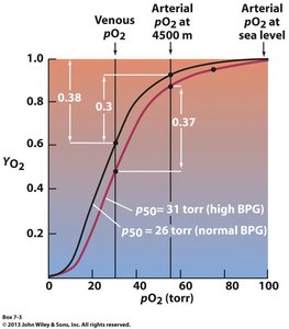
p50: For hemoglobin, p50 is 26 torr.
Hill Equation: , where n is the Hill coefficient (n ≈ 2.8–3.0 for hemoglobin).
Hill Plot: Used to determine the degree of cooperativity.
Mechanism of Cooperative Binding
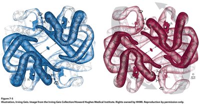
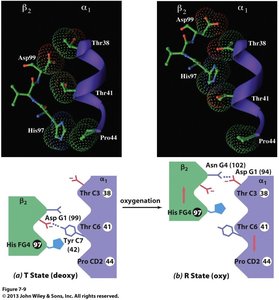
Oxygen binding to one subunit of hemoglobin induces conformational changes that increase the affinity of the remaining subunits for oxygen. This is explained by the T (tense) and R (relaxed) state model:
T State: Low affinity for oxygen; stabilized by salt bridges and hydrogen bonds.
R State: High affinity for oxygen; salt bridges are broken, and the heme iron moves into the plane of the porphyrin ring.
Allosteric Regulation: The transition between T and R states is an example of allosteric regulation.
Bohr Effect and Regulation by CO2 and pH
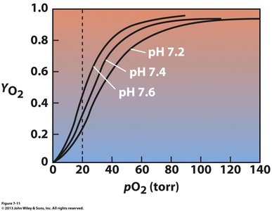
The Bohr effect describes the influence of pH and CO2 on hemoglobin's oxygen affinity:
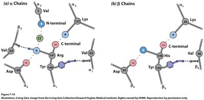
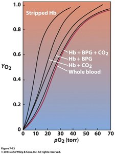
Bohr Effect: Lower pH (higher [H+]) and higher CO2 concentration decrease hemoglobin's affinity for oxygen, promoting O2 release in tissues.
Mechanism: Protonation of certain residues stabilizes the T state; CO2 forms carbamates with N-terminal amino groups, further stabilizing the T state.
Equation:
Regulation by 2,3-Bisphosphoglycerate (BPG)
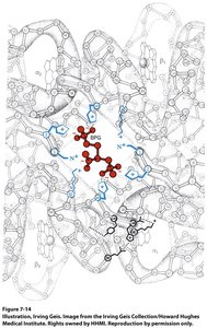
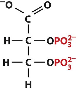
BPG is an allosteric effector that binds to the central cavity of deoxyhemoglobin, stabilizing the T state and reducing oxygen affinity. This is crucial for oxygen delivery under varying physiological conditions, such as high altitude.
BPG Binding: BPG forms hydrogen bonds and ion pairs with the β subunits' N-terminal groups.
Effect: Shifts the oxygen dissociation curve to the right, facilitating oxygen release in tissues.
Adaptation: Increased BPG levels at high altitude decrease hemoglobin's oxygen affinity, enhancing tissue oxygenation.
Protein Function: Antibodies (Immunoglobulins)
Immune System Overview
The immune system protects the body from pathogens through cellular and humoral immunity. Antibodies, or immunoglobulins, are produced by B lymphocytes and recognize specific antigens, marking them for destruction.
Cellular Immunity: Mediated by T cells, targeting virally infected cells.
Humoral Immunity: Mediated by antibodies produced by B cells, effective against bacteria and extracellular pathogens.
Antigen: Any foreign macromolecule (often protein or carbohydrate) that triggers an immune response.
Antibody Structure and Classes
Antibodies are Y-shaped molecules composed of two identical light chains and two identical heavy chains, forming a (LH)2 structure. There are five main classes of immunoglobulins (IgA, IgD, IgE, IgG, IgM), each with distinct functions and heavy chain types.
IgA: Found in mucosal areas; prevents pathogen adherence.
IgD: Function not well understood; may play a role in antimicrobial defense.
IgE: Protects against parasites; involved in allergic responses.
IgG: Most abundant in blood and extracellular fluid; provides long-term immunity.
IgM: First antibody produced in response to infection; forms pentamers.
Antigen-Antibody Interaction and Specificity
Antigen binding occurs at the variable regions of the antibody, specifically within hypervariable sequences (complementarity-determining regions, CDRs). The strength and specificity of binding depend on structural complementarity and are mediated by noncovalent interactions (hydrogen bonds, van der Waals, ionic, and hydrophobic interactions).
Dissociation Constants: Range from 10-4 to 10-10 M, indicating high affinity.
Cross-Linking: Most antibodies are divalent, allowing them to bind two identical antigens and cross-link them, enhancing immune response.
Antibody Diversity and Immune Response
Antibody diversity is generated through somatic recombination and hypermutation of immunoglobulin genes in B cells. Upon antigen exposure, specific B cells proliferate and differentiate into plasma cells (antibody-secreting) and memory cells (long-lived, rapid responders).
Somatic Recombination: Gene segments are rearranged to create diverse antibodies.
Somatic Hypermutation: Point mutations fine-tune antigen specificity.
Monoclonal Antibodies: Produced by hybridoma cells (fusion of myeloma and lymphocyte), used in research and therapy.
Autoimmune Diseases
Normally, the immune system does not react against self-antigens. Loss of tolerance can lead to autoimmune diseases, where antibodies target the body's own molecules.
Summary Table: Comparison of Myoglobin and Hemoglobin
Property | Myoglobin | Hemoglobin |
|---|---|---|
Location | Muscle tissue | Red blood cells |
Structure | Monomer (1 polypeptide) | Tetramer (2α, 2β subunits) |
Heme Groups | 1 | 4 |
O2 Binding Curve | Hyperbolic | Sigmoidal (cooperative) |
Function | O2 storage | O2 transport |
p50 | 2.8 torr | 26 torr |
