 뒤로
뒤로Protein Purification and Characterization Techniques
스터디 가이드 - 스마트 노트
 자료에 맞춘 맞춤형 노트, 핵심 정의, 예시, 맥락을 확장해 제공합니다.
Protein Purification and Characterization
Introduction to Protein Purification
Protein purification is a fundamental process in biochemistry, allowing researchers to isolate a specific protein from complex biological mixtures for further study. The process involves several steps, each exploiting different physical or chemical properties of proteins, such as solubility, charge, size, and binding affinity.

Goal: Obtain pure, active protein for research or therapeutic use.
Challenge: Large amounts of biological material may yield only small quantities of pure protein.
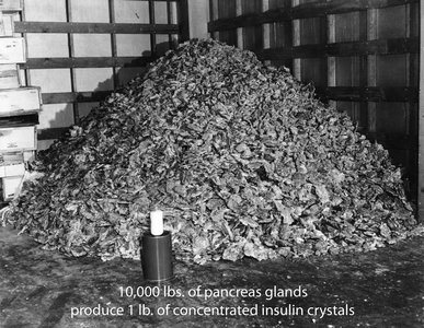
Example: Historically, 10,000 lbs of pancreas glands were required to produce just 1 lb of concentrated insulin crystals.
Initial Protein Extraction and Fractionation
Cell Disruption and Homogenization
To begin purification, cells or tissues are disrupted to release their contents. Homogenization breaks open cells, producing a mixture called a homogenate.
Methods: Mechanical disruption, sonication, enzymatic digestion.
Differential Centrifugation
Differential centrifugation separates cellular components based on size and density by spinning homogenates at increasing speeds.
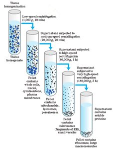
Low-speed centrifugation: Pellets nuclei and unbroken cells.
Medium-speed centrifugation: Pellets mitochondria, lysosomes, peroxisomes.
High-speed centrifugation: Pellets microsomes and small vesicles.
Very high-speed centrifugation: Pellets ribosomes, large macromolecules.
Density Gradient Centrifugation
This technique separates components based on their buoyant density using a gradient (e.g., sucrose). Each component migrates to its equilibrium position in the gradient.
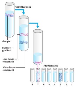
Application: Isolation of organelles, nucleic acids, or protein complexes.
Protein Separation by Solubility
Salting In and Salting Out
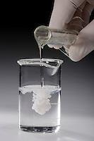
Proteins can be separated based on their solubility in salt solutions. At low salt concentrations, solubility increases (salting in), but at high concentrations, proteins precipitate (salting out).
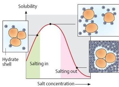
Common salt: Ammonium sulfate ((NH4)2SO4).
Principle: Different proteins precipitate at different salt concentrations, allowing selective separation.
Dialysis
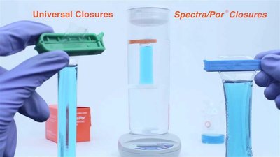
Dialysis removes small molecules (e.g., salts, solvents) from protein solutions by diffusion through a semipermeable membrane, retaining larger protein molecules.
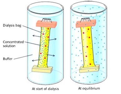
Application: Buffer exchange, desalting after salting out.
Chromatographic Techniques for Protein Purification
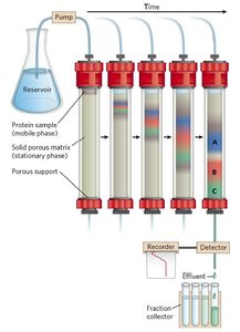
Column Chromatography Overview
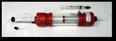
Chromatography separates proteins based on their differential interactions with a stationary phase (solid matrix) and a mobile phase (liquid buffer).
Types: Ion exchange, size exclusion, affinity, and reverse phase chromatography.
Ion Exchange Chromatography
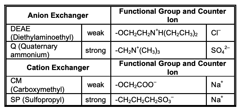
This method separates proteins based on their net charge at a given pH. The column contains charged resin beads that bind proteins of opposite charge.
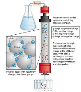
Cation exchangers: Negatively charged resins bind positively charged proteins.
Anion exchangers: Positively charged resins bind negatively charged proteins.
Elution: Proteins are eluted by increasing salt concentration or changing pH.
Example: At pH where peptide A is -3 and peptide B is +1, peptide A elutes first from a cation exchanger, peptide B elutes first from an anion exchanger.
Size Exclusion (Gel Filtration) Chromatography
This technique separates proteins based on size. The column is packed with porous beads; small proteins enter the pores and elute later, while large proteins are excluded and elute earlier.
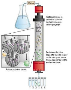
Application: Estimation of molecular weight, removal of aggregates.
Affinity Chromatography
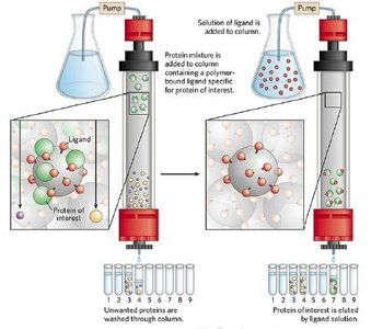
Affinity chromatography exploits specific binding interactions between a protein and a ligand attached to the column matrix. Only proteins with affinity for the ligand are retained and later eluted with a solution containing free ligand or by changing buffer conditions.
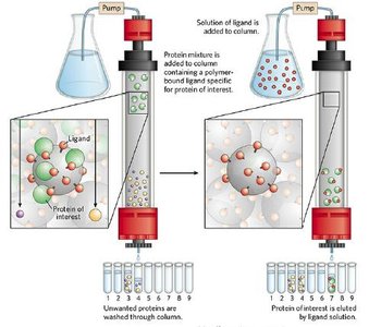
Example: His-tagged proteins bind to nickel columns and are eluted with imidazole.
Assessing Purification: Purification Table
Purification Table and Calculations
Progress of purification is tracked using a purification table, which lists total protein, total activity, specific activity, purification factor, and yield at each step.
Step | Fraction Volume (mL) | Total Protein (mg) | Activity (units) | Specific Activity (units/mg) |
|---|---|---|---|---|
Crude extract | 1400 | 10,000 | 100,000 | 10 |
Ammonium sulfate | 280 | 3,000 | 96,000 | 32 |
Ion exchange | 90 | 400 | 80,000 | 200 |
Size exclusion | 80 | 100 | 60,000 | 600 |
Affinity | 6 | 3 | 45,000 | 15,000 |
Purification factor:
Percent yield:
Protein Characterization by Electrophoresis
Polyacrylamide Gel Electrophoresis (PAGE)
Electrophoresis separates proteins based on their movement in an electric field. Polyacrylamide gels are commonly used for protein separation.
Factors affecting migration: Charge, size, and shape of the protein.
SDS-PAGE
SDS-PAGE is a denaturing electrophoresis technique where proteins are coated with sodium dodecyl sulfate (SDS), giving them a uniform negative charge and allowing separation based on size alone.
SDS: Binds to proteins, denatures them, and imparts a constant charge-to-mass ratio.
Reducing agents: (e.g., β-mercaptoethanol) break disulfide bonds for complete denaturation.
Isoelectric Focusing
Isoelectric focusing separates proteins based on their isoelectric point (pI), the pH at which the protein has no net charge and does not migrate in an electric field.
Application: Determining pI, resolving proteins with similar sizes but different charges.
Two-Dimensional Gel Electrophoresis
This advanced technique combines isoelectric focusing and SDS-PAGE, allowing separation of proteins by both pI and molecular weight. It is highly effective for proteomics studies.
Application: Comparing protein expression in different cell types or conditions (e.g., healthy vs. cancerous cells).
