 뒤로
Protein Secondary Structure: Principles, Types, and Structural Features
스터디 가이드 - 스마트 노트
 자료에 맞춘 맞춤형 노트, 핵심 정의, 예시, 맥락을 확장해 제공합니다.
Protein 3D Structure
Levels of Protein Structure
Proteins exhibit a hierarchical organization of structure, which is essential for their biological function. The four levels of protein structure are:
Primary Structure: The linear sequence of amino acids linked by peptide bonds.
Secondary Structure: Local spatial arrangement of the polypeptide backbone, stabilized by hydrogen bonds. Includes α-helices, β-sheets, loops, and turns.
Tertiary Structure: The overall three-dimensional folding of a single polypeptide chain, determined by interactions among secondary structural elements.
Quaternary Structure: The arrangement of multiple polypeptide chains into a functional protein complex.
Restraints on Local Protein Structure
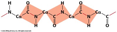
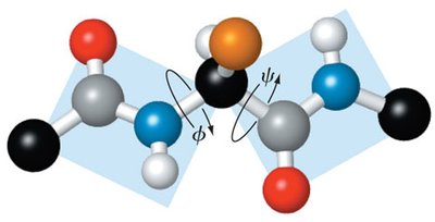
The peptide bond is planar and primarily adopts the trans configuration, restricting rotation. Free rotation is limited to the bonds between the α-carbon and the amide nitrogen (ϕ, phi) and between the α-carbon and the carbonyl carbon (ψ, psi). These are known as dihedral or torsion angles.
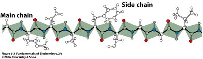
Steric clash: Occurs between backbone and side chain atoms, further limiting possible conformations.
Peptide bond planarity: Ensures that the backbone adopts a relatively rigid structure.
Ramachandran Plot
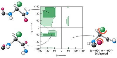
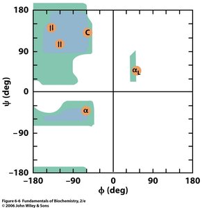
The Ramachandran plot is a graphical representation of the allowed and disallowed conformations of the backbone dihedral angles (ϕ and ψ) for each amino acid in a peptide. Steric clashes define large disallowed regions, while favored regions correspond to common secondary structures.
Allowed regions: Indicate sterically favorable conformations.
Secondary structure correlation: The favored regions correspond to α-helices and β-sheets.
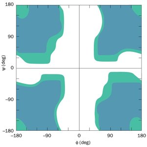
Ramachandran Plot: Glycine
Glycine, due to its small side chain, is allowed across more of the dihedral space than other amino acids, making it more flexible in protein structures.
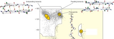
Ramachandran Plot: Secondary Structure
Repeated similar ϕ and ψ angles give rise to secondary structure. Common secondary structures include α helix, parallel β sheet, and antiparallel β sheet.
Secondary Structure Elements
α-Helix
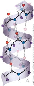
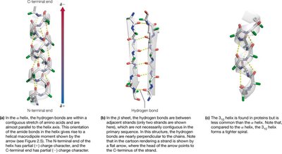
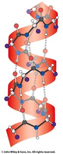
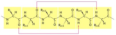
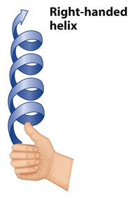
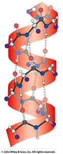
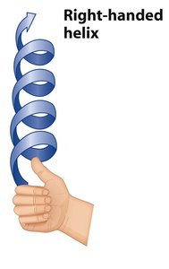
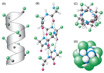
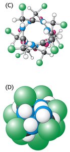
The α-helix is a right-handed helical structure stabilized by hydrogen bonds between the backbone amide and carbonyl groups. It is characterized by specific dihedral angles and a regular hydrogen bonding pattern.
Dihedral angles: ϕ = -57°, ψ = -47°
Residues per turn: 3.6
Pitch: 5.4 Å (distance helix rises per turn)
Hydrogen bonding: C=O of residue i and N-H of residue i+4
Hydrogen bond distance: 2.8 Å
Side chains: Point outward and backward, minimizing steric clashes
Helical Wheel Views and Heptad Repeats
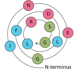
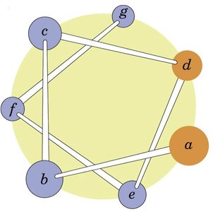
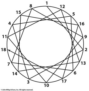
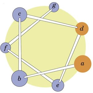
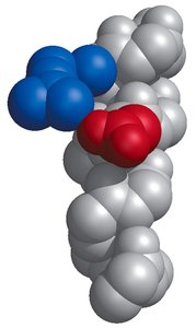
Helical wheel diagrams are used to visualize the spatial arrangement of amino acids in an α-helix, particularly to identify amphipathic helices and heptad repeats (patterns of seven residues).
Sequence Rules for α-Helix Formation
Helical propensity: Linear, non-β-branched amino acids are common in helices; proline and glycine are rare due to steric reasons.
Electrostatics: Similar charges do not cluster, but opposite charges may interact.
Interactions: Amino acids at positions i and i+3 or i+4 are close in space and often associate through ionic or hydrophobic interactions.
Nonstandard Helices
Differences in hydrogen bond patterning can lead to helices with differing numbers of residues per turn, such as the 310 helix.
β-Sheet
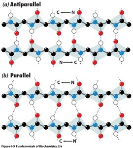
β-sheets are formed by extended polypeptide chains arranged side by side, stabilized by hydrogen bonds between backbone amides and carbonyls. They can be parallel or antiparallel, with distinct hydrogen bonding geometries.
Dihedral angles: Parallel: ϕ = -119°, ψ = 113°; Antiparallel: ϕ = -139°, ψ = 135°
Side chains: Project alternately above and below the plane of the sheet
Hydrogen bonds: Linear in antiparallel sheets, non-linear in parallel sheets
Mixed and Extended β-Sheets
In three dimensions, extended β-sheets are often twisted, and arrows are used to indicate N to C directionality.
Irregular Structure: Turns and Loops
Turns and loops are irregular or nonstandard secondary structural units that connect more standard units. β-hairpin turns generally connect antiparallel β-strands and are stabilized by hydrogen bonds between the first and fourth residues. Glycine and proline are common in turn regions.
Random coil: No detectable structure, high flexibility
Coil structure: Structured regions not fitting standard secondary structure definitions
β-turns: Stabilized by hydrogen bond from the 1st to the 4th residue; proline often at position 2, glycine at position 3
Hydrogen Bonding in Secondary Structure
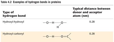
Hydrogen bonds are critical for stabilizing secondary structures. The typical distance between donor and acceptor atoms in protein hydrogen bonds is about 0.28 nm.
Type of hydrogen bond | Typical distance between donor and acceptor atom (nm) |
|---|---|
Hydroxyl-hydroxyl | 0.28 |
Hydroxyl-carbonyl | 0.28 |
Summary: Secondary Structure
Secondary structure is defined by local interactions of the primary sequence, primarily through hydrogen bonding.
α-helices and β-sheets are the main types of secondary structure, each with characteristic dihedral angles and hydrogen bonding patterns.
Turns and loops provide flexibility and connect standard secondary structural elements.
Ramachandran plots are essential for understanding the allowed conformations of protein backbone dihedral angles.
Additional info: The notes have been expanded to provide academic context and explanations for each structural feature, including definitions, examples, and relevant diagrams.
