 뒤로
A Tour of the Cell: Structure and Function of Eukaryotic Cells
스터디 가이드 - 스마트 노트
 자료에 맞춘 맞춤형 노트, 핵심 정의, 예시, 맥락을 확장해 제공합니다.
Cell Structure Overview
Introduction to Cell Structure
Eukaryotic cells are highly organized, containing various membrane-bound organelles and structural components that perform specialized functions. Understanding these structures is essential for grasping cellular processes and their roles in life.
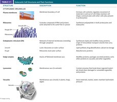
Plasma Membrane: The outer boundary of the cell, composed of a phospholipid bilayer, acts as a selective barrier for the passage of oxygen, nutrients, and waste.
Organelles: Internal structures with specific functions, often surrounded by membranes.

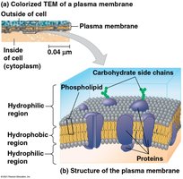
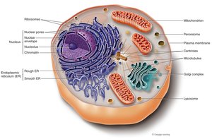
Phospholipid Membrane
Structure and Function
The phospholipid membrane is fundamental to cellular compartmentalization and function.
Phospholipid Bilayer: Composed of hydrophilic heads and hydrophobic tails, forming a semi-permeable barrier.
Functions:
Exchange of membrane material
Transfer of material between organelles
Localization of cellular activities
Work surface for biochemical reactions
Quarantine of reactive compounds
Nucleus and Nuclear Envelope
Composition and Function
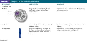
The nucleus is the control center of the cell, housing genetic material and coordinating cellular activities.
Nuclear Envelope: Double membrane separating nuclear material from cytoplasm; contains nuclear pores for material exchange.
Nuclear Matrix: Filamentous protein network organizing chromosomes and maintaining nucleus shape.
Nucleolus: Site of ribosome subunit assembly; synthesizes rRNA and imports ribosomal proteins.
Chromatin: Complex of DNA and proteins; stores genetic information for protein and RNA synthesis.
Cytoplasmic Structures
Ribosomes
Ribosomes are the sites of protein synthesis, composed of RNA and protein.
Free Ribosomes: Suspended in cytosol; synthesize proteins for use within the cell.
Bound Ribosomes: Attached to rough ER; synthesize proteins for export or membrane insertion.
Endomembrane System
Components and Functions
The endomembrane system is a group of interconnected membranous structures that facilitate transport and processing of cellular materials.
Components: Nuclear envelope, endoplasmic reticulum (ER), Golgi apparatus, lysosomes, vacuoles, plasma membrane.
Functions:
Direct connections and vesicle-mediated transport
Modification, sorting, and packaging of molecules
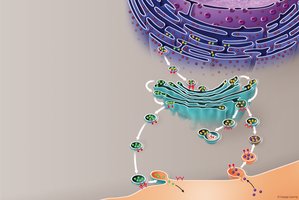
Endoplasmic Reticulum (ER)
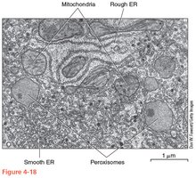
Rough ER (RER): Studded with ribosomes; synthesizes and modifies proteins, adds carbohydrates and lipids, folds proteins with chaperones.
Smooth ER (SER): Lacks ribosomes; synthesizes and modifies lipids, metabolizes carbohydrates, stores calcium ions, detoxifies drugs.
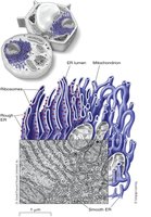
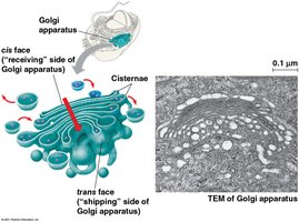
Golgi Complex
Structure: Stacks of flattened membranous sacs (cisternae).
Function: Modifies molecules from ER, sorts and packages materials into vesicles for transport, manufactures certain macromolecules.
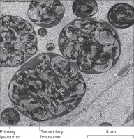
Lysosomes
Contain digestive enzymes: Break down large molecules, autophagy of worn-out cell material, digestion of foreign material.
Types:
Primary: Buds from Golgi
Secondary: Fusion with vesicles
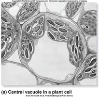
Vacuoles
Types and Functions
Vacuoles are membrane-enclosed sacs with diverse roles in storage, digestion, and water regulation.
Central Vacuole (Plants): Stores water, nutrients, pigments, and waste; maintains turgor pressure for structural support.
Food Vacuole: In animal cells and protists; digests food by fusing with lysosomes.
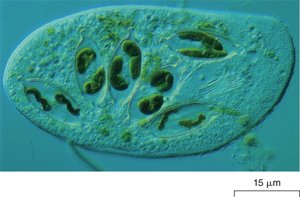
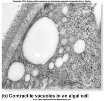
Contractile Vacuole: Removes excess water in freshwater protists.
Peroxisomes
Structure and Function
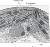
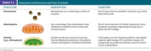
Peroxisomes are small organelles containing enzymes for metabolic reactions, including fatty acid digestion and detoxification.
Functions:
Break down fatty acids
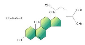
Produce hormones (cholesterol)
Detoxify toxins (e.g., alcohol in liver)
Convert fats to sugars in plant seeds
Byproduct: Hydrogen peroxide (H2O2), decomposed by catalase:
Energy Generators
Mitochondria
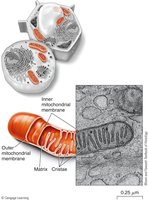
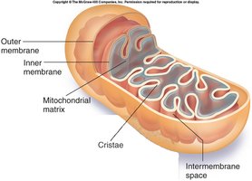
Mitochondria are the powerhouses of the cell, converting food into ATP through aerobic respiration.
Structure: Double membrane, inner membrane folded into cristae, intermembrane space, matrix, circular DNA.
Functions: ATP production, apoptosis (programmed cell death).
Chloroplast
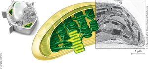
Chloroplasts are found in plant cells and are responsible for photosynthesis, converting light energy into chemical energy.
Components: Circular DNA, thylakoids (light-absorbing pigments: chlorophyll, carotenoids), stroma.
Function: Production of glucose from light energy.
Cytoskeleton
Types and Functions
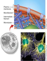
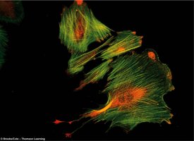
The cytoskeleton is a dense network of protein fibers that provide structural support, motility, and organization within the cell.
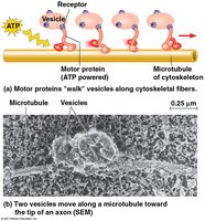
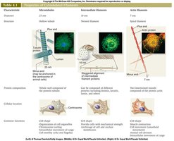
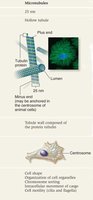
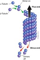
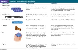
Microtubules: Largest filaments, composed of alpha and beta tubulin dimers; involved in cell shape, chromosome movement, and cell mobility.
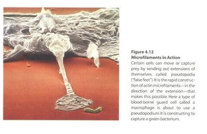
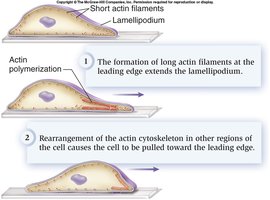
Microfilaments: Most slender, composed of actin; involved in cell movement, structural support, and contractile functions.
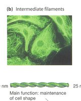
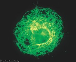
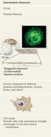
Intermediate Filaments: Intermediate size, composed of various proteins; provide stability and maintain cell shape.
Microfilaments
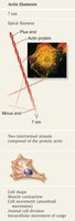
Structure: Two intertwined chains of actin; associated with myosin for contractile functions.
Functions: Cell movement, structural support, formation of cortex, capturing prey.

Intermediate Filaments
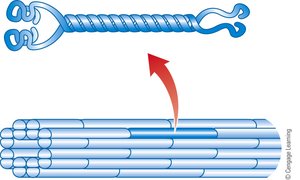
Structure: Twisted strands of various proteins (e.g., keratin, nuclear lamins).
Functions: Stabilize nucleus and organelles, maintain cell shape, provide mechanical strength.
Microtubules
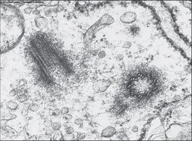
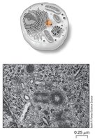
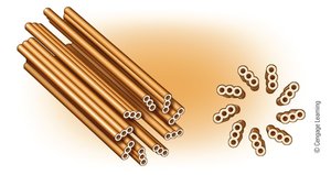
Structure: Hollow tubes of tubulin dimers; anchored in microtubule-organizing centers (MTOC), such as centrosomes.
Functions: Structural support, chromosome movement, cell mobility, formation of cilia and flagella.
Cilia and Flagella
Structure and Function
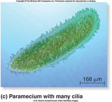
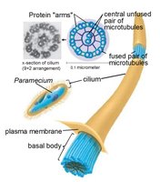
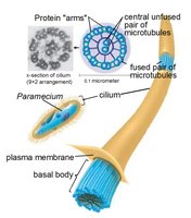
Cilia and flagella are microtubular extensions that provide motility to cells and move materials across cell surfaces.
Cilia: Short, hair-like structures; move in unison to propel cells or move material.
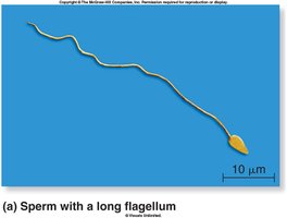
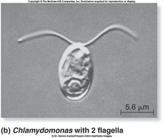
Flagella: Long, tail-like structures; usually singular or few per cell, provide locomotion.
Basal Body: Anchors cilia and flagella; derived from centrioles.
Arrangement: 9+2 microtubule pattern in cilia/flagella; 9+0 in basal bodies.
Movement: Sliding of microtubule pairs powered by dynein proteins.
Extracellular Components
Cell Wall (Plants)
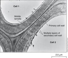
The cell wall provides structural strength, limits water absorption, and protects plant cells.
Composition: Cellulose and lignin; gives rigidity and strength (wood).
Extracellular Matrix (ECM) in Animals
The ECM surrounds animal cells, providing structural support and facilitating cell communication.
Composition: Glycoproteins (mainly collagen and fibronectins), integrins for cell attachment.
Glycocalyx: Allows cell recognition, contact, and adhesion; contributes to tissue strength.
Summary Table: Eukaryotic Cell Structures and Functions
Major Organelles and Their Functions
Structure | Description | Function |
|---|---|---|
Nucleus | Double membrane, contains nucleolus and chromatin | Stores genetic information, coordinates cell activities |
Ribosome | RNA and protein, free or bound | Protein synthesis |
ER (Rough/Smooth) | Membranous network, rough with ribosomes | Protein and lipid synthesis, detoxification |
Golgi Apparatus | Stacked cisternae | Modification, sorting, packaging of molecules |
Lysosome | Membrane sac with enzymes | Intracellular digestion |
Vacuole | Membrane sac | Storage, water regulation |
Peroxisome | Enzyme-containing sac | Metabolic reactions, detoxification |
Mitochondria | Double membrane, cristae | ATP production |
Chloroplast | Double membrane, thylakoids | Photosynthesis |
Cytoskeleton | Microtubules, microfilaments, intermediate filaments | Support, motility, organization |
Organization of the Cell
Coordination of Cell Activities
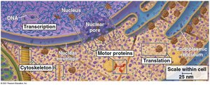
The organization of cellular structures enables efficient coordination of cell activities, including transcription, translation, and transport.
Motor Proteins: Facilitate movement of vesicles and organelles along cytoskeletal tracks.
Compartmentalization: Allows simultaneous and specialized cellular processes.
Conclusion
Summary
Eukaryotic cells are complex and highly organized, with specialized structures that perform essential functions for life. Understanding these components is fundamental to the study of biology and cellular processes. Additional info: Some details and explanations were expanded for academic completeness and clarity.
