 뒤로
A Tour of the Cell: Structure, Function, and Organization
스터디 가이드 - 스마트 노트
 자료에 맞춘 맞춤형 노트, 핵심 정의, 예시, 맥락을 확장해 제공합니다.
Introduction to the Cell
Discovery and Importance of Cells
Cells are the fundamental units of life, discovered with the advent of microscopes in the 1600s. Understanding cell structure and function is essential for all biological sciences.
Cell Theory: All living things are composed of cells; the cell is the structural and functional unit of life; all cells arise from preexisting cells.
Microscopes: Essential tools for visualizing cells and their components.
Microscopy and Cell Visualization
Types of Microscopes
Microscopes allow scientists to observe cells and their structures at various magnifications and resolutions.
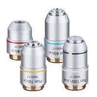
Light Microscope (LM): Uses light to magnify images; suitable for viewing living cells.
Electron Microscope (EM): Uses electron beams for higher magnification and resolution; only dead specimens, no color.
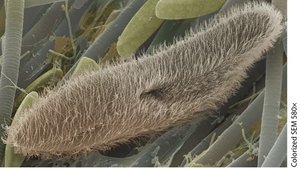
SEM (Scanning Electron Microscope): Provides detailed 3D images of cell surfaces.
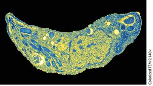
TEM (Transmission Electron Microscope): Reveals internal cell structures in 2D.
Magnification and Resolution
Magnification is the increase in an object's image size compared to its actual size, while resolution is the clarity of the image.
Magnification: Product of ocular and objective lens powers.
Resolution: Higher in electron microscopes than light microscopes.
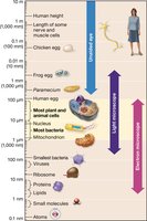
Scale of Biological Structures
Cells and their components vary greatly in size, from atoms to entire organisms.
Cell Size and Surface Area-to-Volume Ratio
Why Cells Are Small
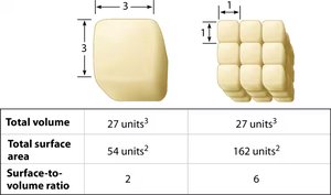
Cell size is limited by the surface area-to-volume ratio, which affects nutrient and waste exchange efficiency.
High Surface Area to Low Volume Ratio: Ideal for efficient exchange.
Large Cells: Less efficient; tend to divide into smaller cells.
Cell Membrane Structure
Plasma Membrane Function
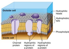
The plasma membrane separates the cell's internal environment from the external environment, regulating the movement of substances.
Structure: Composed of a phospholipid bilayer with embedded proteins.
Function: Controls exchange, maintains homeostasis.
Prokaryotic Cell Structure
Shared Components of All Cells
Both prokaryotic and eukaryotic cells share certain fundamental structures.
Plasma/Cell Membrane
Cytoplasm
Genetic Material (DNA)
Ribosomes
Prokaryotic External Structures
Prokaryotic cells have unique external features that may be optional.
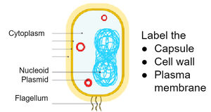
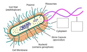
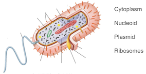
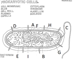
Capsule: Jellylike coating, sometimes pathogenic.
Cell Wall: Rigid structure outside the membrane.
Flagella: Long, thin structures for movement.
Fimbrae: Attachment structures.
Prokaryotic Internal Structures
Internal components of prokaryotic cells include:
Cytoplasm: Site of most reactions.
Ribosomes: Site of protein synthesis.
Nucleoid: Area containing main DNA.
Plasmid: Optional ring of extra DNA.
Eukaryotic Cell Structure
Nucleus and Ribosomes
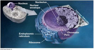
The nucleus is the defining feature of eukaryotic cells, safeguarding genetic material and regulating access.
Nuclear Envelope: Double membrane system.
Nuclear Pores: Allow selective transport.
Nucleoplasm: Fluid suspending DNA.
Nucleolus: Ribosome subunit production.
The Endomembrane System
Components and Functions
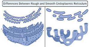
The endomembrane system is a network of membranes for producing, modifying, and transporting proteins and lipids.
Endoplasmic Reticulum (ER): Rough ER (with ribosomes) makes proteins; Smooth ER (no ribosomes) makes lipids and detoxifies.
Golgi Apparatus: Modifies, packages, and distributes cell products.
Vesicles: Transport, store, and dispose of materials.
Lysosomes: Digest cell debris and waste.
Peroxisomes: Break down fatty acids and toxins.
Vacuoles: Storage; contractile vacuoles expel excess water, central vacuoles in plants store water and nutrients.
Energy-Converting Organelles
Mitochondria and Chloroplasts
Mitochondria and chloroplasts are specialized organelles for energy conversion.
Mitochondria: Site of cellular respiration; have two membranes, their own DNA and ribosomes.
Chloroplasts: Site of photosynthesis in plants and some protists; have three membranes, stroma, and thylakoids.
Endosymbiont Theory: Suggests mitochondria and chloroplasts originated from prokaryotes living inside larger cells.
The Cytoskeleton and Cell Surfaces
Cytoskeleton Structure and Function
The cytoskeleton is a network of protein filaments that provides structural support, movement, and organization within the cell.
Microtubules: Hollow tubes for movement, division, and internal transport; grow from the centrosome.
Cilia and Flagella: Specialized microtubule structures for locomotion.
Intermediate Filaments: Reinforce cell shape and anchor organelles.
Microfilaments: Solid rods of actin for support and movement.
Extracellular Matrix and Cell Walls
Some cells have external structures for support and communication.
Extracellular Matrix: Holds cells together, supports plasma membrane, attaches to integrins.
Plant Cell Wall: Provides protection and skeletal support; plasmodesmata connect adjacent cells.
Summary Table: Prokaryotic Cell Structures
Structure | Function |
|---|---|
Capsule | Adhesion, protection |
Cell Wall | Structural support |
Cell Membrane | Regulates exchange |
Flagellum | Movement |
Fimbrae | Attachment |
Nucleoid | Main genetic material |
Plasmid | Extra DNA, benefits |
Cytoplasm | Site of reactions |
Ribosome | Protein synthesis |
Key Equations
Surface Area of a Cube:
Volume of a Cube:
Surface Area-to-Volume Ratio:
Review Questions
Describe the importance of microscopes in understanding cell structure and function.
Distinguish between prokaryotic and eukaryotic cell structures.
Explain how cell size is limited by surface area-to-volume ratio.
Compare the structures and functions of mitochondria and chloroplasts.
Describe the evidence for the endosymbiont theory.
Compare microfilaments, intermediate filaments, and microtubules.
Relate the structure of cilia and flagella to their functions.
Describe the four functional categories of organelles in eukaryotic cells.
