 뒤로
뒤로Axial Skeleton Study Guide: Bone Features, Skull, and Vertebral Column
스터디 가이드 - 스마트 노트
 자료에 맞춘 맞춤형 노트, 핵심 정의, 예시, 맥락을 확장해 제공합니다.
Q1. Define the anatomical features of bones (Articulations, Extensions, Depressions, Passages)
Background
Topic: Bone Markings and Features
This question tests your understanding of the terminology used to describe the shapes, projections, depressions, and openings found on bones. These terms are foundational for identifying and describing skeletal structures in anatomy.
Key Terms and Definitions:
Articulations: Where two bones meet (e.g., condyle, facet, head).
Extensions and Projections: Raised or elongated parts of bones (e.g., crest, epicondyle, line, process, trochanter, tubercle, tuberosity, spine).
Depressions: Indentations or holes in bones (e.g., alveolus, fossa, fovea, sulcus/groove).
Passages: Openings for nerves and blood vessels (e.g., canal/meatus, foramen, fissure).
Step-by-Step Guidance
Review each term and try to recall or look up its definition in your textbook or lecture notes.
Group the terms by their category (articulation, projection, depression, passage) to help remember their function and location.
Write a brief definition for each term, focusing on its anatomical significance (e.g., a condyle is a rounded articular projection).
Think of examples of each feature on actual bones (e.g., the head of the femur, the foramen magnum in the skull).
Try solving on your own before revealing the answer!
Final Answer:
Condyle: Rounded articular projection.
Facet: Smooth, nearly flat articular surface.
Head: Bony expansion carried on a narrow neck.
Crest: Narrow, prominent ridge of bone.
Epicondyle: Raised area on or above a condyle.
Line: Narrow ridge of bone; less prominent than a crest.
Process: Any bony prominence.
Trochanter: Large, blunt, irregular surface (femur only).
Tubercle: Small, rounded projection or process.
Tuberosity: Large, rounded projection; may be roughened.
Spine: Sharp, slender, often pointed projection.
Alveolus: Deep pit or socket (e.g., teeth in jaw).
Fossa: Shallow, basinlike depression.
Fovea: Small pit or depression.
Sulcus (groove): Furrow or groove.
Canal (meatus): Tubelike passageway.
Foramen (foramina): Round or oval opening through a bone.
Fissure: Narrow, slitlike opening.
These definitions are essential for describing bone anatomy and will help you identify features on skeletal diagrams and models.
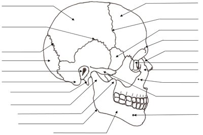
Q2. Label the specific structures at the end of each arrow in the lateral view of the skull (image_1)
Background
Topic: Skull Anatomy
This question tests your ability to identify the major bones and features of the skull from a lateral (side) view. Recognizing these structures is crucial for understanding cranial anatomy and the relationships between different bones.
Key Terms and Structures:
Common bones to identify: frontal, parietal, occipital, temporal, sphenoid, zygomatic, maxilla, mandible, nasal, lacrimal.
Key features: mastoid process, styloid process, external auditory meatus, coronal suture, squamous suture, lambdoid suture, alveolar processes.
Step-by-Step Guidance
Start by identifying the largest bones (e.g., parietal, frontal, occipital) and their general locations.
Locate the major sutures (coronal, squamous, lambdoid) that separate these bones.
Identify the facial bones (maxilla, mandible, zygomatic, nasal) and their positions relative to the cranial bones.
Look for distinctive features such as the mastoid process (behind the ear), the external auditory meatus (ear canal), and the zygomatic arch (cheekbone).
Label each arrow with the correct anatomical term based on its location and shape.
Try solving on your own before revealing the answer!
Final Answer:
Common labels (from top to bottom, left to right):
Parietal bone
Frontal bone
Occipital bone
Temporal bone
Sphenoid bone
Nasal bone
Lacrimal bone
Zygomatic bone
Maxilla
Mandible
Mastoid process
Styloid process
Coronal suture
Squamous suture
Lambdoid suture
External auditory meatus
Alveolar processes (maxilla and mandible)
Check your diagram against these terms to ensure you can identify each structure on the skull.
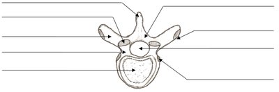
Q3. Label the specific structures at the end of each arrow in the vertebra diagram (image_2)
Background
Topic: Vertebral Anatomy
This question tests your ability to identify the main features of a typical vertebra, which is essential for understanding the structure and function of the vertebral column.
Key Terms and Structures:
Body (centrum)
Vertebral foramen
Spinous process
Transverse process
Superior articular process
Inferior articular process
Lamina
Pedicle
Step-by-Step Guidance
Identify the large, rounded anterior portion as the vertebral body.
Locate the central hole (vertebral foramen) through which the spinal cord passes.
Find the posterior projection (spinous process) and the lateral projections (transverse processes).
Identify the superior and inferior articular processes, which articulate with adjacent vertebrae.
Label the lamina and pedicle, which form the vertebral arch.
Try solving on your own before revealing the answer!
Final Answer:
Body (centrum)
Vertebral foramen
Spinous process
Transverse process
Superior articular process
Inferior articular process
Lamina
Pedicle
These are the main anatomical features of a typical vertebra. Practice labeling them on diagrams and models for mastery.
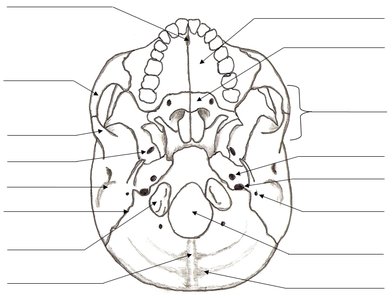
Q4. Label the specific structures at the end of each arrow in the inferior view of the skull (image_3)
Background
Topic: Inferior Skull Anatomy
This question tests your ability to identify the bones and foramina visible from the underside of the skull. This view is important for understanding the passage of nerves and blood vessels into and out of the cranial cavity.
Key Terms and Structures:
Foramen magnum
Occipital condyles
Palatine bone
Vomer
Maxilla
Hard palate
Jugular foramen
Carotid canal
Styloid process
Mastoid process
Sphenoid bone
Foramen ovale, spinosum, lacerum
Step-by-Step Guidance
Locate the large central opening (foramen magnum) and the occipital condyles on either side.
Identify the hard palate (formed by the maxilla and palatine bones) at the anterior end.
Find the vomer (part of the nasal septum) and the palatine bone.
Locate the various foramina (openings) for nerves and vessels, such as the jugular foramen and carotid canal.
Label the styloid and mastoid processes, as well as the sphenoid bone and its associated foramina.
Try solving on your own before revealing the answer!
Final Answer:
Foramen magnum
Occipital condyles
Palatine bone
Vomer
Maxilla
Hard palate
Jugular foramen
Carotid canal
Styloid process
Mastoid process
Sphenoid bone
Foramen ovale, spinosum, lacerum
Review the labeled diagram to reinforce your understanding of the inferior skull anatomy.
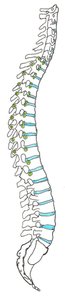
Q5. Locate and label the following structures or regions on the vertebral column diagram (image_4): Intervertebral discs, spinous process, costal facet, intervertebral foramen, coccyx, sacrum, auricular surface of sacrum, transverse process, lumbar vertebra, lumbar curvature, cervical vertebra, cervical curvature, thoracic vertebra, thoracic curvature, sacral curvature.
Background
Topic: Vertebral Column Anatomy
This question tests your ability to identify the major regions and features of the vertebral column, including the different types of vertebrae, curvatures, and specific anatomical landmarks.
Key Terms and Structures:
Intervertebral discs
Spinous process
Costal facet
Intervertebral foramen
Coccyx
Sacrum
Auricular surface of sacrum
Transverse process
Lumbar vertebra
Lumbar curvature
Cervical vertebra
Cervical curvature
Thoracic vertebra
Thoracic curvature
Sacral curvature
Step-by-Step Guidance
Identify the four main regions of the vertebral column: cervical, thoracic, lumbar, and sacral (including coccyx).
Locate the curvatures: cervical (neck), thoracic (upper back), lumbar (lower back), sacral (pelvic region).
Find the intervertebral discs between the vertebrae and the spinous and transverse processes projecting from each vertebra.
Locate the sacrum and coccyx at the base of the column, and identify the auricular surface of the sacrum.
Label the costal facets (on thoracic vertebrae) and intervertebral foramina (spaces for spinal nerves).
Try solving on your own before revealing the answer!
Final Answer:
Cervical vertebrae (top 7)
Cervical curvature (neck region)
Thoracic vertebrae (next 12)
Thoracic curvature (upper/mid back)
Lumbar vertebrae (next 5)
Lumbar curvature (lower back)
Sacrum (5 fused vertebrae)
Sacral curvature (pelvic region)
Coccyx (tailbone)
Intervertebral discs (between vertebrae)
Spinous process (posterior projection)
Transverse process (lateral projection)
Costal facet (on thoracic vertebrae)
Intervertebral foramen (lateral openings for nerves)
Auricular surface of sacrum (articulates with pelvis)
Use this labeled diagram to practice identifying each structure and region on the vertebral column.
