 뒤로
뒤로Cell Communication and Signaling: Mechanisms and Examples
스터디 가이드 - 스마트 노트
 자료에 맞춘 맞춤형 노트, 핵심 정의, 예시, 맥락을 확장해 제공합니다.
Cell Communication and Signaling
Overview of Cell Signaling
Cell signaling is a fundamental process by which cells receive, interpret, and respond to external and internal signals. This process enables multicellular organisms to coordinate activities such as growth, metabolism, and responses to environmental changes.
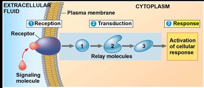
External signals are converted into cellular responses through a series of steps.
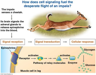
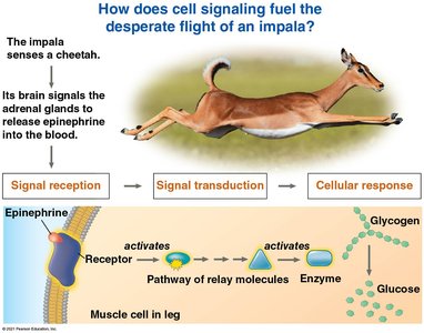
Epinephrine (adrenaline) is a hormone released during stress, stimulating the breakdown of glycogen in liver and muscle cells.
Cell signaling can be local (adjacent cells) or long-distance (hormones traveling through the bloodstream).
Local Signaling: Direct Contact
Cells can communicate directly through physical contact, which is essential for development and immune responses.
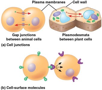
Cell junctions connect the cytoplasm of adjacent cells:
Plasmodesmata (plants): channels for water, solutes, proteins, and RNA.
Gap junctions (animals): cytoplasmic channels between cells.
Cell–cell recognition: membrane-bound molecules on one cell bind to another, important in embryonic development and immunity.
Local Signaling: Secreted Messengers
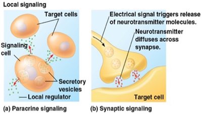
Some cells communicate by releasing signaling molecules that affect nearby target cells.
Paracrine signaling: local regulators (e.g., growth factors) stimulate nearby cells.
Synaptic signaling: neurons release neurotransmitters across synapses to target cells.
Long-Distance Signaling: Hormones
Hormones are used for long-distance communication in both plants and animals.
Endocrine signaling: hormones released into the bloodstream reach distant target cells.
Only cells with specific receptors respond to a given hormone.
Classes of Chemical Messengers
Chemical messengers are classified by their structure and solubility, which determines their transport and receptor location.
Peptide/Protein: Chains of amino acids, water-soluble, act on cell-surface receptors.
Steroid: Lipids built from cholesterol, lipid-soluble, act on intracellular receptors.
Amine: Modified amino acids (e.g., epinephrine, thyroid hormone), solubility varies.
Solubility determines:
How messengers move in the blood
Where their receptors are located
The type and speed of cellular response
The Three Stages of Cell Signaling
Reception, Transduction, and Response
Cell signaling involves three main stages: reception, transduction, and response. Each stage is essential for converting an external signal into a functional cellular change.
Reception: A signaling molecule (ligand) binds to a receptor protein.
Transduction: The receptor changes shape and initiates a signal transduction pathway.
Response: The transduced signal triggers a specific cellular response.
Reset: Components are switched off so the cell can respond to new signals.
Signal Reception: Types of Receptors
Receptors are proteins that bind signaling molecules and initiate cellular responses. Their location and mechanism depend on the type of messenger.
G Protein-Coupled Receptors (GPCRs): Bind hormones and neurotransmitters; largest family of cell-surface receptors.
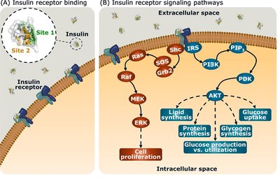
Receptor Tyrosine Kinases (RTKs): Bind growth factors and insulin; catalyze phosphorylation.
Ion Channel Receptors: Ligand-gated channels for ions; rapid changes in ion concentration.
Intracellular Receptors: Located in cytoplasm or nucleus; bind steroid and thyroid hormones.
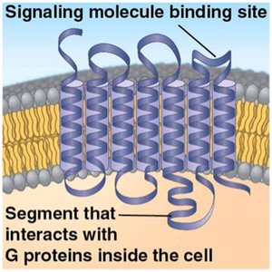
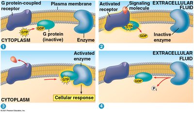
G Protein-Coupled Receptors (GPCRs)
GPCRs are transmembrane proteins that transmit signals via G proteins, which act as molecular switches.
Inactive G protein binds GDP.
Signal binding activates GPCR, GTP replaces GDP, G protein becomes active.
Active G protein activates an enzyme, leading to a cellular response.
Reset: G protein hydrolyzes GTP to GDP, becomes inactive.
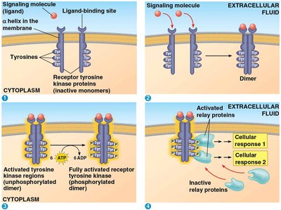
Receptor Tyrosine Kinases (RTKs)
RTKs are membrane receptors that transfer phosphate groups from ATP to proteins, often triggering multiple pathways.
Ligands include growth factors.
RTKs can activate several signal transduction pathways simultaneously.
Abnormal RTK function is linked to cancers.
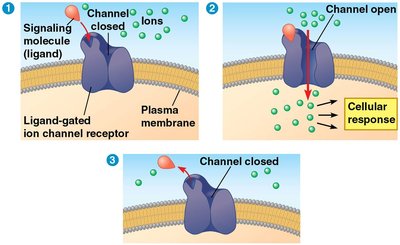
Ligand-Gated Ion Channel Receptors
These receptors act as gates for ions, opening or closing in response to ligand binding.
Ligand binding opens the channel, allowing ions (e.g., Na⁺, Ca²⁺) to flow.
Rapid changes in ion concentration trigger cellular responses.
Channel closes when ligand leaves.
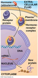
Intracellular Receptors
Intracellular receptors bind small or hydrophobic messengers that cross the plasma membrane, such as steroid and thyroid hormones.
The hormone–receptor complex acts as a transcription factor, regulating gene expression.
Example: Aldosterone regulates salt and water balance.
Signal Transduction Pathways
Phosphorylation Cascades
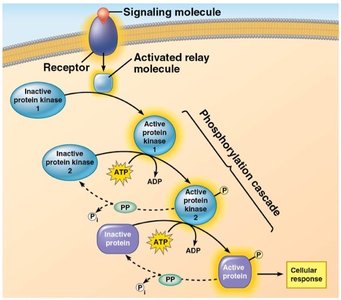
Signal transduction often involves cascades of protein kinases, which relay and amplify signals by phosphorylation.
Each activated protein activates the next in the pathway.
Protein kinases add phosphate groups; protein phosphatases remove them.
This acts as a molecular switch for cellular activities.
Second Messengers
Second messengers are small, nonprotein molecules that spread signals within the cell.
Common second messengers: cyclic AMP (cAMP), calcium ions (Ca²⁺).
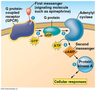
Adenylyl cyclase converts ATP to cAMP; phosphodiesterase converts cAMP to AMP.

cAMP in G Protein Pathways
cAMP is a key second messenger in pathways initiated by GPCRs.
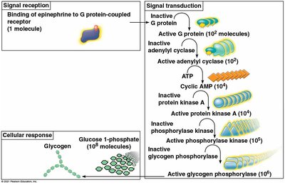
First messenger binds GPCR → activates G protein → activates adenylyl cyclase → produces cAMP → activates protein kinase A → triggers cellular responses.
Cellular Responses
Nuclear Responses
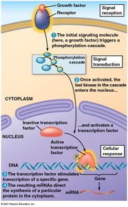
Cell signaling can regulate gene expression by activating transcription factors in the nucleus.
Pathways often turn genes on or off, controlling protein synthesis.
Example: Growth factors activate specific genes.
Cytoplasmic Responses
Signaling can also trigger responses in the cytoplasm, such as the breakdown of glycogen.
Epinephrine stimulates glycogen breakdown, releasing glucose for energy.
Signal Amplification
Signal amplification allows a small number of signaling molecules to produce a large cellular response.
Each step in the pathway amplifies the signal.
Example: One epinephrine molecule can lead to the production of millions of glucose molecules.
Specificity of Cellular Responses
Only cells with the appropriate receptor respond to a specific messenger. Different cells may respond differently to the same signal.
Messenger and receptor fit together like a key and lock.
Different collections of proteins in cells lead to varied responses.
Resetting the Signal: Termination
To respond to new signals, activated components must be switched off.
Ligand leaves receptor; G protein hydrolyzes GTP; cAMP is removed; proteins are dephosphorylated; ion channels close.
The cell returns to its resting state, ready for new signals.
Case Study: Epinephrine Signaling in Muscle Cells
Flight Response in Animals
When an impala senses a predator, its brain signals the adrenal glands to release epinephrine, triggering a cascade that mobilizes energy for rapid movement.
Reception: Epinephrine binds to GPCR on muscle cells.
Transduction: G protein → adenylyl cyclase → cAMP → protein kinase A → kinase cascade.
Response: Glycogen phosphorylase breaks down glycogen into glucose-1-phosphate.
Reset: Signal components are deactivated.
Insulin Signaling and Diabetes
Role of Insulin
Insulin is a peptide hormone that facilitates glucose uptake into cells, where it is stored as glycogen. Diabetes results from insufficient insulin production or improper insulin use.
Insulin binds to its receptor, triggering a cascade that allows glucose to enter cells.
Hydrolysis of glycogen releases glucose when needed for energy.
Review Questions
Which would cause the most problems for a cell-signaling pathway involving a G protein-coupled receptor?
A steroid hormone is bound by an intracellular receptor. The resulting complex is most likely to do what?
Which gives the most complete and correct description of a signal transduction pathway?
There is a lack of ATP present as a signal transduction cascade begins to form. How is this process most likely going to be affected?
Summary Table: Classes of Chemical Messengers
Class | Examples | Structure | Solubility | Transport in Blood | Receptor Location |
|---|---|---|---|---|---|
Peptide/Protein | Insulin, glucagon, oxytocin | Chain of amino acids | Water-soluble (hydrophilic) | Dissolves freely in plasma | Plasma membrane (GPCR, RTK) |
Steroid | Aldosterone, cortisol, estrogen, testosterone | Lipid built from cholesterol (four fused rings) | Lipid-soluble (hydrophobic) | Bound to carrier proteins | Intracellular (cytoplasm or nucleus) |
Amine | Epinephrine, norepinephrine, thyroid hormone | Modified single amino acid | Epinephrine: water-soluble; Thyroid hormone: lipid-soluble | Epinephrine: free in plasma; Thyroid hormone: on carrier proteins | Epinephrine: membrane GPCR; Thyroid hormone: nuclear receptor |
Additional info: These notes expand on the original lecture slides and images, providing definitions, examples, and context for each concept. The summary table is inferred from the content and organizes key properties of messenger classes for clarity.
