 뒤로
뒤로Cell Cycle, Mitosis, and Meiosis: Structured Study Notes
스터디 가이드 - 스마트 노트
 자료에 맞춘 맞춤형 노트, 핵심 정의, 예시, 맥락을 확장해 제공합니다.
The Cell Cycle
Overview: The Key Roles of Cell Division
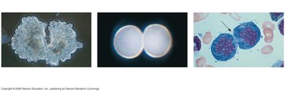
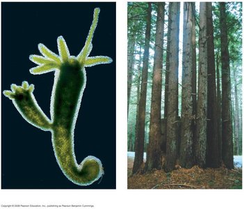
Cell division is fundamental to life, enabling reproduction, growth, and repair in organisms. The cell cycle encompasses the life of a cell from its formation to its own division.
Unicellular organisms reproduce by dividing a single cell.
Multicellular organisms rely on cell division for development, growth, and tissue renewal.
Cell division produces genetically identical daughter cells through mitosis, or genetically diverse cells through meiosis.
Cellular Organization of Genetic Material
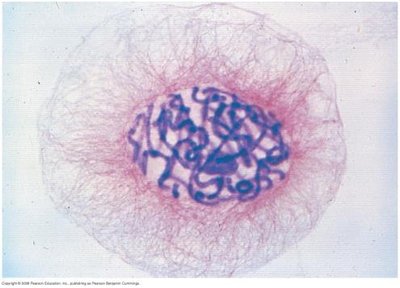
The genetic material of a cell is organized into chromosomes, which are composed of DNA and proteins (chromatin). Chromatin condenses into chromosomes during cell division.
Genome: All DNA in a cell.
Prokaryotes: Single DNA molecule.
Eukaryotes: Multiple DNA molecules.
Chromosome Duplication and Separation
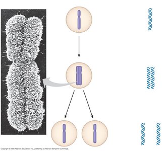
Before cell division, chromosomes are duplicated, forming two sister chromatids joined at the centromere. During division, sister chromatids separate, ensuring each daughter cell receives identical genetic information.
Phases of the Cell Cycle
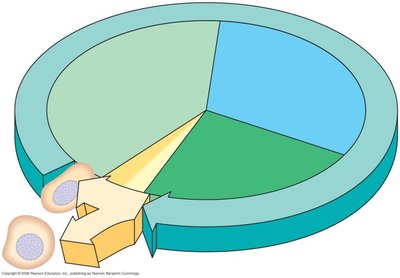
The cell cycle consists of interphase (G1, S, G2) and the mitotic phase (mitosis and cytokinesis).
G1 phase: Cell growth and organelle development.
S phase: DNA synthesis and chromosome duplication.
G2 phase: Further growth and preparation for division.
M phase: Mitosis (nuclear division) and cytokinesis (cytoplasmic division).
Mitosis
Phases of Mitosis
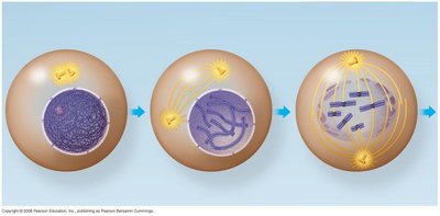
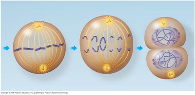
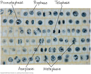
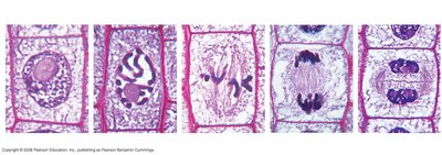
Mitosis is divided into five phases: prophase, prometaphase, metaphase, anaphase, and telophase, followed by cytokinesis.
Prophase: Chromatin condenses, nucleoli disappear, centrosomes move apart.
Prometaphase: Nuclear envelope breaks down, spindle microtubules attach to kinetochores.
Metaphase: Chromosomes align at the metaphase plate.
Anaphase: Sister chromatids separate and move to opposite poles.
Telophase: Nuclei reform, chromosomes decondense.
Cytokinesis: Cytoplasm divides, forming two daughter cells.
The Mitotic Spindle
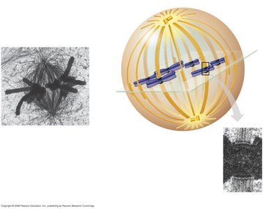
The mitotic spindle is a structure of microtubules that orchestrates chromosome movement during mitosis. It includes centrosomes, spindle microtubules, and asters.
Spindle microtubules attach to kinetochores during prometaphase.
Chromosomes align at the metaphase plate.
Microtubules shorten during anaphase, separating chromatids.
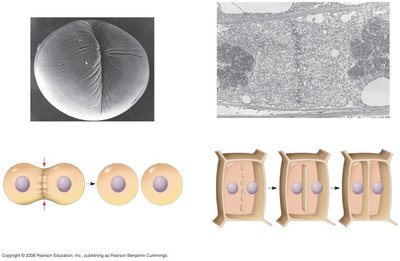
Cytokinesis: Animal vs. Plant Cells
Cytokinesis differs between animal and plant cells:
Animal cells: Cleavage furrow forms, pinching the cell in two.
Plant cells: Cell plate forms, leading to a new cell wall between daughter cells.
Binary Fission
Cell Division in Prokaryotes
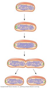
Prokaryotes reproduce by binary fission, a process where the chromosome replicates and daughter chromosomes move apart.
Evolution of Mitosis
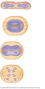
Mitosis likely evolved from binary fission. Some protists display intermediate forms of cell division.
Regulation of the Cell Cycle
Cell Cycle Control System
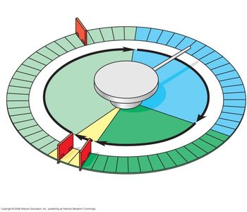
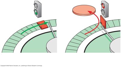
The cell cycle is regulated by a molecular control system, with checkpoints ensuring proper progression.
G1 checkpoint: Most important; determines if cell proceeds to division.
G2 and M checkpoints: Ensure readiness for mitosis and chromosome separation.
Cells not receiving signals enter G0, a non-dividing state.
Cyclins and Cyclin-Dependent Kinases (Cdks)
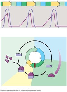
Cyclins and Cdks are regulatory proteins controlling cell cycle progression. MPF (maturation-promoting factor) is a cyclin-Cdk complex that triggers entry into M phase.
Internal and External Signals
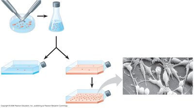
Cell division is regulated by internal signals (e.g., unattached kinetochores delay anaphase) and external signals (e.g., growth factors like PDGF stimulate division).
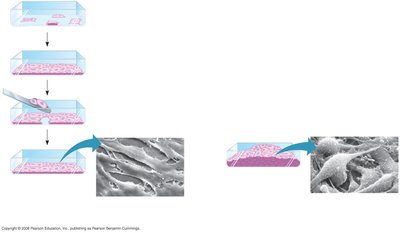
Loss of Cell Cycle Controls in Cancer Cells
Cancer cells evade normal cell cycle controls, leading to uncontrolled growth and tumor formation. Tumors can be benign, malignant, or metastatic.
Benign: Remain at original site.
Malignant: Invade surrounding tissues.
Metastatic: Spread to other parts of the body.
Meiosis and Sexual Life Cycles
Genetics and Heredity
Genetics is the study of heredity and variation. Genes, located at specific loci on chromosomes, are inherited through gametes.
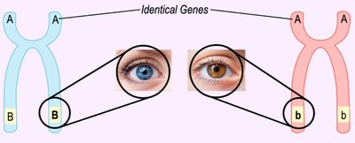
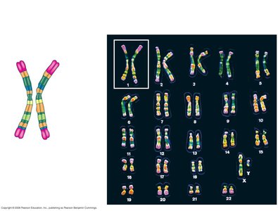
Chromosome Sets in Human Cells
Human somatic cells have 23 pairs of chromosomes (46 total), including autosomes and sex chromosomes. Gametes are haploid (n = 23).
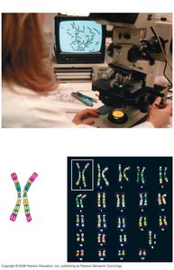
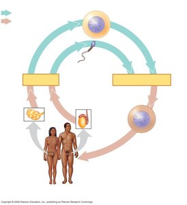
Sexual Life Cycles
Sexual reproduction alternates between meiosis and fertilization, maintaining chromosome number across generations. Three main life cycle types exist in animals, plants, fungi, and protists.
Meiosis
Reduction of Chromosome Number
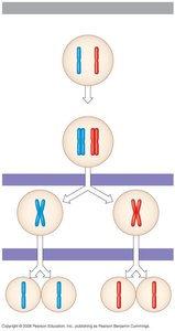
Meiosis reduces chromosome number from diploid (2n) to haploid (n) through two consecutive divisions: meiosis I and meiosis II.
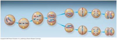
Stages of Meiosis
Meiosis I: Prophase I, Metaphase I, Anaphase I, Telophase I and cytokinesis. Meiosis II: Prophase II, Metaphase II, Anaphase II, Telophase II and cytokinesis.
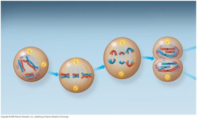
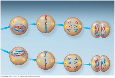
Comparison of Mitosis and Meiosis
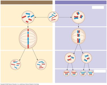
Mitosis produces genetically identical diploid cells; meiosis produces genetically diverse haploid cells. Three events unique to meiosis I: synapsis and crossing over, tetrad formation at metaphase plate, separation of homologous chromosomes.
Genetic Variation in Sexual Life Cycles
Mechanisms of Genetic Variation
Three mechanisms contribute to genetic variation:
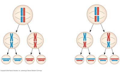
Independent assortment: Random orientation of homologous chromosomes during meiosis I.
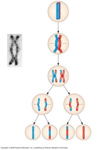
Crossing over: Exchange of genetic material between nonsister chromatids during prophase I.
Random fertilization: Any sperm can fertilize any egg, increasing genetic diversity.
Evolutionary Significance
Genetic variation is essential for evolution, as it provides the raw material for natural selection.
Summary Table: Mitosis vs. Meiosis
Property | Mitosis | Meiosis |
|---|---|---|
DNA Replication | Occurs during interphase before mitosis | Occurs during interphase before meiosis I |
Number of Divisions | One | Two |
Synapsis of Homologous Chromosomes | Does not occur | Occurs during prophase I |
Number of Daughter Cells | Two, diploid, genetically identical | Four, haploid, genetically diverse |
Role in Animal Body | Growth, repair, asexual reproduction | Produces gametes, introduces genetic variability |
Additional info: Cohesins are protein complexes responsible for sister chromatid cohesion, cleaved at different stages in mitosis and meiosis.
