 뒤로
뒤로Cell Membrane Structure and Function: Mini-Textbook Study Guide
스터디 가이드 - 스마트 노트
 자료에 맞춘 맞춤형 노트, 핵심 정의, 예시, 맥락을 확장해 제공합니다.
The Cell Membrane
Functions of the Cell Membrane
The cell membrane is a fundamental structure that separates the living cell from its environment, forming a stable boundary between two aqueous compartments. It performs a variety of essential functions:
Selective uptake and export: Controls the movement of ions and molecules in and out of the cell.
Compartmentalization: Maintains distinct internal environments within the cell.
Protein sorting: Directs proteins to their proper locations.
Anchoring cytoskeleton: Provides structural support and shape.
Production of energy intermediates: Generates ATP and NADPH.
Cell signaling: Facilitates communication between cells.
Cell and nuclear division: Participates in mitosis and meiosis.
Adhesion: Enables cells to stick to each other and to the extracellular matrix.
Components of the Cell Membrane
The cell membrane consists of two major components:
Phospholipids: Form the phospholipid bilayer, the basic structural framework.
Proteins: Embedded within the bilayer, performing specialized functions.
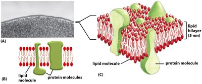
Phospholipid Bilayer
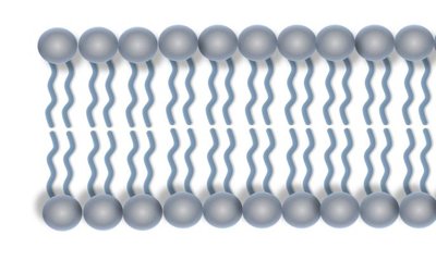
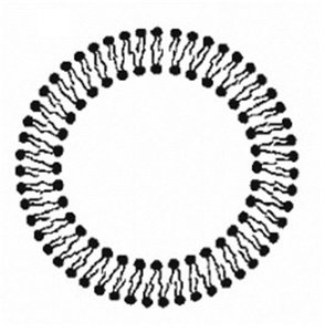
Phospholipids are amphipathic molecules with a hydrophilic (water-attracting) head and two hydrophobic (water-repelling) tails. When placed in water, they self-assemble into a bilayer, with hydrophobic tails facing inward and hydrophilic heads facing outward.
The Fluid Mosaic Model
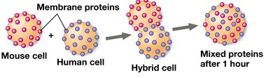
The cell membrane is described by the fluid mosaic model, which states that the membrane is a dynamic structure with various molecules (phospholipids, proteins, cholesterol, carbohydrates) moving freely within the plane of the membrane.
Types of Phospholipid Movements
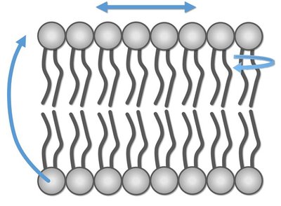
Phospholipids exhibit several types of movement within the membrane:
Lateral movement: Phospholipids move side-to-side within the same layer.
Rotation: Phospholipids spin in place.
Flip-flop: Rarely, phospholipids switch layers, but this is energetically unfavorable due to hydrophobic interactions.
Membrane Fluidity and Its Regulation
Factors Affecting Membrane Fluidity
Membrane fluidity is influenced by several factors:
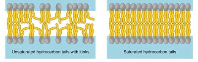
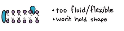
Degree of unsaturation: Unsaturated fatty acids have double bonds, causing kinks that increase spacing and fluidity.
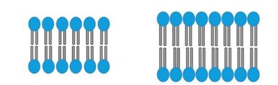
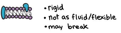
Fatty acid length: Longer tails increase intermolecular interactions, reducing fluidity.
Temperature: Higher temperatures increase fluidity; lower temperatures decrease it.
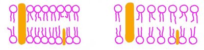
Cholesterol: Acts as a fluidity buffer, decreasing fluidity at high temperatures and increasing it at low temperatures.
Degree of Unsaturation
Unsaturated hydrocarbon tails with kinks prevent tight packing, increasing membrane fluidity. Saturated tails pack closely, decreasing fluidity.
Fatty Acid Length
Shorter fatty acid tails result in fewer interactions and greater fluidity, while longer tails promote rigidity.
Temperature and Cholesterol
Cholesterol modulates membrane fluidity depending on temperature, preventing membranes from becoming too rigid or too fluid.
Selective Permeability of the Membrane
What Can Cross the Membrane?
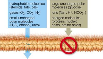
The cell membrane is selectively permeable, allowing some molecules to pass while blocking others. Permeability depends on size and polarity:
Small, nonpolar molecules: Can cross easily (e.g., O2, CO2).
Small, polar uncharged molecules: May cross slowly (e.g., H2O).
Large or charged molecules: Cannot cross without assistance (e.g., glucose, ions).
Transport Across the Membrane
Concentration Gradient and Diffusion
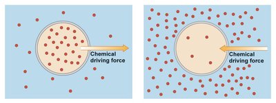
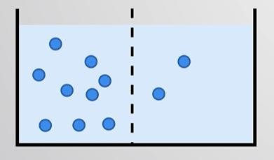
A concentration gradient is a difference in the concentration of a substance between two areas. Diffusion is the tendency for molecules to spread out evenly, moving from high to low concentration until equilibrium is reached.
Gradient Directionality
Movement down the concentration gradient (high to low) is passive and does not require energy. Movement up the gradient (low to high) is active and requires energy.
Electrical and Electrochemical Gradients
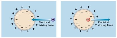
An electrical gradient (membrane potential) is a separation of charge across a membrane. The electrochemical gradient combines both concentration and electrical gradients, influencing ion movement.
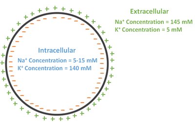
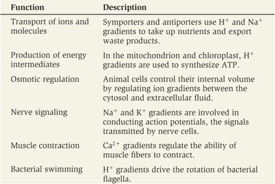
Functions of Ion Electrochemical Gradients
Ion gradients are essential for various cellular functions:
Function | Description |
|---|---|
Transport of ions and molecules | Symporters and antiporters use H+ and Na+ gradients to take up nutrients and export waste products. |
Production of energy intermediates | H+ gradients are used to synthesize ATP in mitochondria and chloroplasts. |
Osmotic regulation | Animal cells regulate ion gradients between cytosol and extracellular fluid. |
Nerve signaling | Na+ and K+ gradients are involved in action potentials. |
Muscle contraction | Ca2+ gradients regulate muscle fiber contraction. |
Bacterial swimming | H+ gradients drive bacterial flagella rotation. |
Types of Membrane Transport
Passive Transport
Passive transport moves substances down their concentration gradient without energy input. Types include:
Simple diffusion: Direct movement through the membrane.
Osmosis: Diffusion of water across a semipermeable membrane.
Facilitated diffusion: Movement through membrane proteins.
Simple Diffusion
Small, nonpolar molecules move directly through the membrane from high to low concentration.
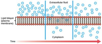
Osmosis
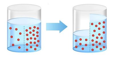
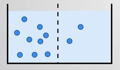
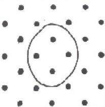
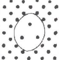
Osmosis is the net diffusion of water toward higher solute concentration. Water moves across the membrane to balance solute concentrations.
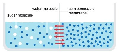
Tonicity
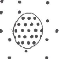
Tonicity describes the effect of a solution on cell volume:
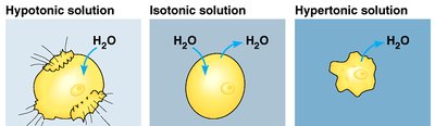
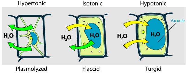
Isotonic: Equal solute concentration inside and outside the cell; no net water movement.
Hypertonic: Higher solute concentration outside; water moves out, cell shrinks.
Hypotonic: Lower solute concentration outside; water moves in, cell swells.
Cells Without a Cell Wall
Animal cells in hypotonic solutions swell and may burst; in hypertonic solutions, they shrink.
Cells With a Cell Wall
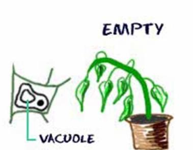
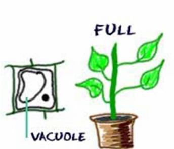
Plant cells in hypotonic solutions become turgid (preferred state); in hypertonic solutions, they undergo plasmolysis.
Membrane Proteins
Types of Membrane Proteins
Membrane proteins are responsible for most specific functions of membranes:
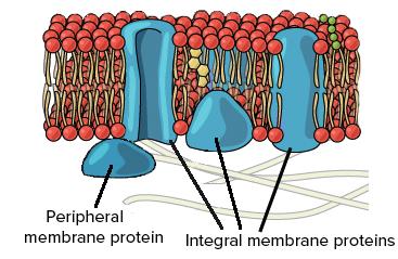
Integral proteins: Penetrate the hydrophobic interior of the bilayer.
Peripheral proteins: Associate with the bilayer but do not penetrate it.
Transmembrane Proteins
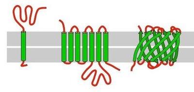
Integral proteins that span the entire membrane width, often involved in transport. Hydrophobic regions interact with the bilayer interior; hydrophilic regions face aqueous environments.
Facilitated Diffusion
Channel and Carrier Proteins
Facilitated diffusion allows molecules that cannot cross the membrane directly to pass through protein channels or carriers. This process does not require energy and does not alter the direction of transport.
Channel Proteins
Leak channels: Always open, allow passive movement (e.g., aquaporins for water).
Ligand-gated channels: Open in response to ligand binding.
Voltage-gated channels: Open in response to changes in membrane potential.
Mechanically-gated channels: Open in response to mechanical stimuli.
Carrier Proteins
Uniporter: Transports one substance.
Symporter: Transports two substances in the same direction.
Antiporter: Transports two substances in opposite directions.
Active Transport
Primary and Secondary Active Transport
Active transport moves substances against their concentration gradient, requiring energy and transmembrane proteins.
Primary active transport: Uses ATP hydrolysis for energy (e.g., Na+/K+ pump).
Secondary active transport: Couples passive movement of one solute to active transport of another.
Bulk Transport
Types of Bulk Transport
Exocytosis: Vesicles fuse with the membrane to release contents outside.
Endocytosis: Membrane engulfs substances to bring them inside.
Phagocytosis: Cell engulfs large particles.
Pinocytosis: Cell takes in fluid and dissolved molecules.
Receptor-mediated endocytosis: Cell acquires specific substances via receptor binding.
Other Functions of the Membrane
Attachment and Intercellular Joining
Attachment to cytoskeleton: Restricts protein mobility, maintains cell shape.
Intercellular joining: Cells adhere and communicate via junctions (tight, desmosomes, gap junctions, plasmodesmata).
Cell Signaling
Stages of Cell Signaling
Reception: Receptor detects signal (ligand binding).
Transduction: Signal converted to cellular response (often via phosphorylation).
Response: Cellular activity triggered.
Types of Cell Surface Receptors
Enzyme-linked receptors: Ligand binding activates intracellular enzyme.
G-protein-coupled receptors: Ligand binding activates G-protein, triggering response.
Ligand-gated ion channels: Ligand binding opens channel for ion passage.
Types of Cell Signaling
Direct contact: Gap junctions and plasmodesmata.
Juxtacrine: Ligand and receptor on cell surfaces; requires contact.
Paracrine: Ligand released to nearby cells.
Synaptic: Specialized paracrine signaling in neurons.
Endocrine: Hormones travel through bloodstream to distant cells.
Autocrine: Cell releases ligand that binds to its own receptor.
Additional info: This study guide expands on lecture notes with definitions, examples, and academic context for General Biology students.
