 뒤로
Cell Signaling, Metabolism, and DNA Replication: Study Notes for General Biology
스터디 가이드 - 스마트 노트
 자료에 맞춘 맞춤형 노트, 핵심 정의, 예시, 맥락을 확장해 제공합니다.
Cell Signaling and Communication
Types of Cell Signaling
Cell signaling is the process by which cells communicate with each other to coordinate biological activities. There are several modes of signaling based on the distance between cells and the nature of the signaling molecules.
Paracrine Signaling: Involves the release of local regulators (e.g., growth factors) that affect nearby cells. These molecules bind to receptors such as receptor tyrosine kinases (RTKs).
Synaptic Signaling: Occurs in the nervous system, where neurotransmitters are released into the synaptic cleft between neurons to stimulate target cells.
Endocrine (Long-Distance) Signaling: Hormones are released into the bloodstream and travel to distant target cells. Examples include insulin, adrenaline, and growth hormone. Lipid-soluble hormones (e.g., testosterone, estrogen) can cross the plasma membrane and bind to intracellular receptors.
Stages of Cell Signaling
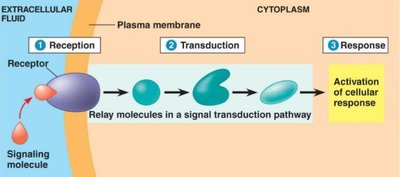
Cell signaling typically involves three main stages: reception, transduction, and response.
Reception: The target cell detects a signaling molecule (ligand) when it binds to a specific receptor protein, usually on the cell surface. The binding causes a conformational change in the receptor, activating it.
Transduction: The activated receptor initiates a cascade of molecular events, often involving phosphorylation and dephosphorylation, which amplifies the signal.
Response: The signal transduction pathway leads to a specific cellular response, such as gene expression or metabolic changes.
Synaptic Signaling Example
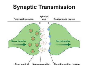
Synaptic transmission is a specialized form of cell signaling in neurons. Neurotransmitters are released from the presynaptic neuron into the synaptic gap and bind to receptors on the postsynaptic neuron, propagating the nerve impulse.
Axon terminal: Releases neurotransmitters.
Neurotransmitter receptor: Receives the signal on the postsynaptic neuron.
Cell Surface and Intracellular Receptors
Receptors can be located on the cell surface or inside the cell. Surface receptors include GPCRs, RTKs, and ligand-gated ion channels. Intracellular receptors are typically engaged by steroid hormones and can act as transcription factors in the nucleus.
GPCRs: Largest family of cell surface receptors, involved in many physiological processes.
RTKs: Bind growth factors and trigger phosphorylation cascades.
Ligand-gated ion channels: Open or close in response to ligand binding, regulating ion flow.
Intracellular receptors: Bind lipid-soluble hormones and regulate gene expression.
Signal Transduction Pathways
Signal transduction often involves phosphorylation cascades mediated by protein kinases and phosphatases. Molecular switches (phosphorylation = ON, dephosphorylation = OFF) regulate the activity of proteins in the pathway.
Protein kinases: Catalyze phosphorylation, activating proteins.
Protein phosphatases: Remove phosphate groups, deactivating proteins.
Second Messengers
Second messengers are small molecules or ions that relay signals inside the cell. Examples include cyclic AMP (cAMP) and calcium ions.
cAMP: Produced by adenylyl cyclase, activates protein kinases.
Calcium ions: Regulate various cellular processes.
G-Protein Coupled Receptor (GPCR) Signaling Pathway
GPCRs are activated by extracellular signaling molecules, leading to a series of intracellular events:
Signaling molecule binds to GPCR, causing a conformational change.
G-protein binds to the receptor with GDP (inactive).
GDP is replaced by GTP (active).
Activated G-protein activates adenylyl cyclase.
Adenylyl cyclase converts ATP to cAMP (second messenger).
cAMP activates protein kinases, leading to phosphorylation cascades.
Introduction to Metabolism
Free Energy Change (ΔG)
Free energy (G) is the portion of a system’s energy available to do work. The change in free energy () determines whether a reaction is spontaneous.
Exergonic reactions: is negative; spontaneous and energy-releasing.
Endergonic reactions: is positive; require energy input.
Equation:
Example: Cellular respiration is exergonic ( kcal/mol), while photosynthesis is endergonic ( kcal/mol).
ATP and Energy Coupling
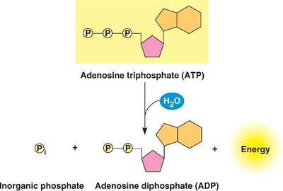
ATP (adenosine triphosphate) powers cellular work by coupling exergonic and endergonic reactions. Hydrolysis of ATP releases energy used for chemical, transport, and mechanical work.
ATP Structure: Adenine, ribose sugar, three phosphate groups.
Hydrolysis Reaction: ATP + H2O → ADP + Pi + Energy
ΔG for ATP hydrolysis: -7.3 kcal/mol (in vitro), -13 kcal/mol (in cell).
Enzymes and Metabolic Reactions
Enzymes are biological catalysts that speed up metabolic reactions by lowering activation energy. Each enzyme is specific to its substrate, determined by the shape of its active site.
Lock and Key Model: Old model; substrate fits exactly into the enzyme.
Induced Fit Model: New model; enzyme changes shape to fit the substrate.
Active Site: Region where substrate binds and reaction occurs.
Factors Influencing Enzyme Activity:
Temperature and pH
Cofactors (ions) and coenzymes (organic molecules)
Enzyme inhibitors (competitive and noncompetitive)
Allosteric regulation (activators and inhibitors)
Cellular Respiration
Overview of Cellular Respiration
Cellular respiration is the process by which cells convert glucose and oxygen into ATP, carbon dioxide, and water. It involves three main stages: glycolysis, citric acid cycle, and electron transport chain.
Catabolic Pathways and ATP Production
Glycolysis: Occurs in the cytoplasm; splits glucose into two pyruvate molecules; net gain of 2 ATP.
Citric Acid Cycle: Occurs in the mitochondrial matrix; breaks down pyruvate; produces CO2, 4 ATP, 10 NADH, 2 FADH2.
Electron Transport Chain: Occurs in the inner mitochondrial membrane; uses NADH and FADH2 to produce 26-28 ATP and water; oxygen is the final electron acceptor.
Table: Stages of Cellular Respiration
Stage | Location | Input | Output |
|---|---|---|---|
Glycolysis | Cytoplasm | Glucose | Net 2 ATP, 2 Pyruvate |
Citric Acid Cycle | Mitochondrial Matrix | 2 Pyruvate | CO2, 4 ATP, 10 NADH, 2 FADH2 |
Electron Transport Chain | Inner Membrane of Mitochondria | 10 NADH, 2 FADH2, O2 | 26-28 ATP, H2O |
Anaerobic Respiration and Fermentation
When oxygen is unavailable, cells use anaerobic respiration or fermentation to produce ATP.
Anaerobic respiration: Uses alternative electron acceptors (e.g., sulfate ions), producing hydrogen sulfide.
Alcohol fermentation: Pyruvate is converted to ethanol and CO2.
Lactic acid fermentation: Pyruvate is converted directly to lactic acid.
DNA Replication
Basic Principles of DNA Replication
DNA replication is semi-conservative, meaning each new DNA molecule consists of one old (template) strand and one new strand. Both prokaryotic and eukaryotic cells follow similar mechanisms, with some differences.
Prokaryotes: Circular DNA, single origin of replication.
Eukaryotes: Linear DNA, multiple origins of replication.
Key Enzymes and Proteins in DNA Replication
DNA Polymerase: Adds nucleotides to the 3’-OH end; DNA Pol III is the main enzyme for synthesis; DNA Pol I removes RNA primers and replaces them with DNA.
Primase: Synthesizes RNA primers to initiate DNA synthesis.
Helicase: Unwinds the DNA helix.
Single-Stranded Binding Proteins (SSBPs): Prevent re-annealing of DNA strands.
Topoisomerase: Relieves tension ahead of the replication fork.
Sliding Clamp: Helps DNA Pol III stay attached to DNA.
DNA Ligase: Seals gaps between Okazaki fragments on the lagging strand.
Leading and Lagging Strand Synthesis
Leading strand: Synthesized continuously towards the replication fork.
Lagging strand: Synthesized discontinuously away from the fork, forming Okazaki fragments.
DNA Pol I: Removes RNA primers and replaces them with DNA.
DNA Ligase: Joins Okazaki fragments.
Additional info: DNA replication is highly regulated and involves proofreading mechanisms to ensure fidelity. DNA Pol III has 3’→5’ exonuclease activity for error correction.
