 뒤로
Cell Theory, Microscopy, and Cell Structure: Foundations of Modern Biology
스터디 가이드 - 스마트 노트
 자료에 맞춘 맞춤형 노트, 핵심 정의, 예시, 맥락을 확장해 제공합니다.
Cell Theory
Historical Development of Cell Theory
The cell theory is a fundamental concept in biology that describes the properties of cells, the basic unit of life. It was developed through the work of several scientists over centuries, beginning with the invention of the microscope.
Robert Hooke (1665): First observed cork cells using a microscope and coined the term "cells" because they resembled small rooms in a monastery.
Anton van Leeuwenhoek (1674): Observed single-celled organisms in pond water, which he called "animalcules." His work expanded the understanding of the diversity of life at the microscopic level.
Matthias Schleiden (1838): Proposed that all plants are made of cells.
Theodor Schwann (1839): Stated that all animals are also made of cells, generalizing the concept to all living things.
Rudolf Virchow (1855): Hypothesized that all cells arise from pre-existing cells, refuting the idea of spontaneous generation.
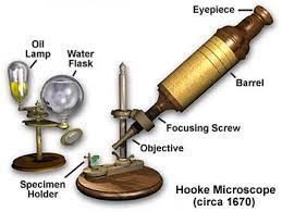
The Three Parts of Cell Theory
All living things are made of cells. This means that cells are the basic building blocks of all organisms, from bacteria to plants and animals.
Cells are the smallest unit of life. While atoms and molecules are smaller, they are not alive. The cell is the smallest structure capable of performing all the functions necessary for life.
All cells come from pre-existing cells. New cells are produced by the division of existing cells, ensuring continuity of life.
Microscopy and the Study of Cells
The Role of Microscopes
The invention and improvement of microscopes enabled scientists to observe cells and their structures, leading to major advances in biology.
Compound Light Microscope: Uses light and multiple lenses to magnify specimens up to 2,000x. Suitable for viewing living cells.
Electron Microscope: Uses beams of electrons for much higher magnification (up to 200,000x), but only for non-living specimens.
Transmission Electron Microscope (TEM): Produces 2D images by passing electrons through thin specimens.
Scanning Electron Microscope (SEM): Produces 3D images by bouncing electrons off the surface of specimens.
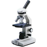
Microscopic Observation of Cells
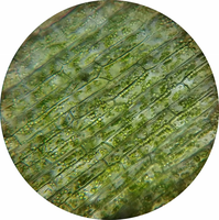
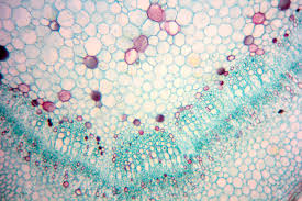
Microscopes reveal the structure and organization of cells, including their organelles and arrangement in tissues.
Plant cells: Typically rectangular due to the presence of a rigid cell wall. Contain chloroplasts, which are visible as green structures under the microscope.
Animal cells: Usually rounder and lack a cell wall and chloroplasts.
Characteristics of Living Things
What Makes Something Alive?
To be considered living, an organism must:
Have DNA
Change over time (evolve)
Use materials and energy
Grow and develop
Respond to the environment
Reproduce
Maintain homeostasis (stable internal conditions)
Cell Size and Organization
Size of Cells
Cells are microscopic, typically about 1/10 the diameter of a human hair. Larger organisms have more cells, not larger cells.
Humans are composed of over 30 trillion cells.
Having many cells allows for specialization and redundancy (backup cells for repair and replacement).
Types of Cells and Organisms
Prokaryotes vs. Eukaryotes
Prokaryotes: Single-celled organisms without a nucleus or membrane-bound organelles. DNA is circular and located in the cytoplasm. Examples: bacteria, blue-green algae.
Eukaryotes: Can be single-celled or multicellular. Have a nucleus and membrane-bound organelles. Examples: plants, animals, fungi, protists.
Similarities Between Prokaryotes and Eukaryotes
Both can be single-celled.
Both may have cilia or flagella for movement.
Cell Structure and Organelles
Major Organelles and Their Functions
Cell Membrane: Phospholipid bilayer that is selectively permeable, controlling what enters and exits the cell.
Cytoplasm: Jelly-like substance filling the cell, containing water, proteins, nucleic acids, minerals, and ions.
Cytoskeleton: Network of protein fibers providing structure, movement, and transport within the cell.
Nucleus: Stores and protects DNA; has pores for molecular exchange.
Nucleolus: Site of ribosome production within the nucleus.
Ribosomes: Synthesize proteins using instructions from DNA.
Endoplasmic Reticulum (ER): Network of membranes producing proteins (rough ER) and lipids (smooth ER); also detoxifies substances.
Golgi Apparatus: Processes, sorts, and ships proteins and lipids.
Vesicles: Membrane-bound sacs for transport within the cell.
Mitochondria: Site of cellular respiration; converts food into usable energy (ATP).
Vacuole: Stores materials; large central vacuole in plants stores water and maintains structure.
Lysosomes: Contain enzymes for digestion and defense (mainly in animal cells).
Peroxisomes: Break down toxic substances like hydrogen peroxide.
Cell Wall: Provides structural support (plants, fungi, some bacteria).
Chloroplasts: Site of photosynthesis in plants and algae.
Cell Transport Mechanisms
Cell Membrane Structure and Function
The cell membrane is a dynamic structure composed of a phospholipid bilayer with embedded proteins, cholesterol, and carbohydrates. It is described by the fluid mosaic model and is selectively permeable.
Types of Transport
Passive Transport: Movement of molecules without energy input. Includes diffusion (movement from high to low concentration) and facilitated diffusion (via protein channels).
Osmosis: Diffusion of water across a membrane through aquaporins. Water moves from areas of high to low concentration.
Active Transport: Movement of molecules against the concentration gradient (low to high), requiring energy (ATP) and protein pumps.
Endocytosis: Cell engulfs large molecules or liquids by folding in the membrane.
Exocytosis: Vesicles fuse with the membrane to release substances outside the cell.
Osmosis and Tonicity
Isotonic Solution: Equal concentration of solutes inside and outside the cell; no net water movement.
Hypertonic Solution: Higher solute concentration outside; water leaves the cell, causing it to shrink.
Hypotonic Solution: Lower solute concentration outside; water enters the cell, which may swell and burst (lyse).
ATP: The Energy Currency
Adenosine Triphosphate (ATP): Organic molecule that stores and provides energy for cellular processes. Produced mainly in mitochondria.
Summary Table: Prokaryotes vs. Eukaryotes
Feature | Prokaryotes | Eukaryotes |
|---|---|---|
Nucleus | No | Yes |
Membrane-bound Organelles | No | Yes |
DNA Structure | Circular | Linear, in nucleus |
Examples | Bacteria, Archaea | Plants, Animals, Fungi, Protists |
