 뒤로
뒤로Cell Types and Microscopy: Prokaryotes, Eukaryotes, and Lab Techniques
스터디 가이드 - 스마트 노트
 자료에 맞춘 맞춤형 노트, 핵심 정의, 예시, 맥락을 확장해 제공합니다.
Cell Types: Prokaryotes vs. Eukaryotes
Overview of Cell Types
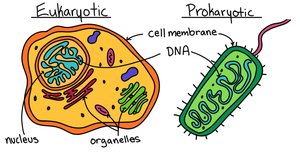
Cells are the fundamental units of life, classified into two main types: prokaryotic and eukaryotic cells. Understanding their structural differences is essential for studying biological diversity and cellular function.
Prokaryotes: Include Bacteria and Archaea. They are unicellular, lack organelles, and do not have a membrane-bound nucleus.
Eukaryotes: Include Protists, Fungi, Plants, and Animals. They can be unicellular or multicellular, possess a membrane-bound nucleus, and contain organelles.
Key Structural Differences
Prokaryotic Cells: DNA is located in the nucleoid region, surrounded by a cell membrane and cell wall. No membrane-bound organelles.
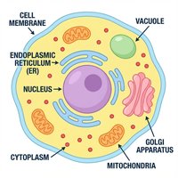
Eukaryotic Cells: DNA is enclosed within a nucleus. Organelles such as mitochondria, endoplasmic reticulum, and Golgi apparatus are present.
Prokaryotes: Bacterial Cell Shapes
Bacterial Morphology
Bacteria are classified based on their shape, which is a key feature for identification and understanding their ecological roles.
Cocci: Spherical-shaped bacteria, often found in clusters or chains.
Bacilli: Rod-shaped bacteria, can occur singly or in chains.
Spirilla: Spiral-shaped bacteria, less common and often motile.
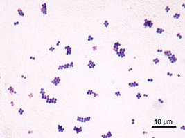
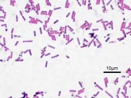
Example: On a mixed slide, all three shapes may be observed together, demonstrating bacterial diversity.
Eukaryotes: Plant Cells
Plant Cell Structure
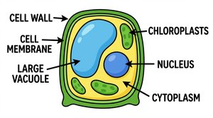
Plant cells are eukaryotic and possess unique features that distinguish them from other cell types.
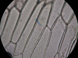
Cell Wall: Provides structural support and protection.
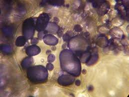
Plastids: Include chloroplasts (photosynthesis), chromoplasts (pigment storage), and amyloplasts (starch storage).
Central Vacuole: Maintains cell turgor and stores nutrients.
Nucleus: Contains genetic material.
Microscopic Observation of Plant Cells
Onion Cells: Used to observe cell walls and nuclei.
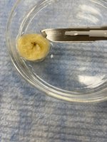
Potato/Banana Cells: Used to observe amyloplasts (starch storage).
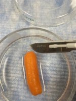
Carrot Cells: Used to observe chromoplasts (pigment storage).
Eukaryotes: Animal Cells
Animal Cell Structure
Animal cells are eukaryotic, multicellular, and lack certain structures found in plant cells.
Cell Membrane: Surrounds the cell, regulating entry and exit of substances.
No Cell Wall: Unlike plant cells, animal cells do not have a rigid cell wall.
No Plastids or Central Vacuole: These organelles are absent in animal cells.
Microscopic Observation of Animal Cells
Frog Blood: Used to observe cell membrane, cytoplasm, and nucleus.
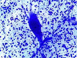
Nerve Tissue: Used to observe neurons and their unique structures.
Eukaryotes: Protists
Protist Cell Structure
Protists are a diverse group of eukaryotes, ranging from single-celled to colonial organisms. They typically have a cell membrane and may lack a cell wall.
Stentor: Ciliated protist, often observed for its unique shape and movement.
Amoeba: Moves using pseudopods, lacks a rigid cell wall.
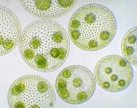
Volvox: Colonial protist, forms spherical colonies with daughter colonies inside.
Microscopy Techniques and Lab Safety
Microscope Operation
Proper use of the microscope is essential for accurate observation and analysis of cells.
Start with the 4X objective lens to locate and center the specimen.
Use coarse adjustment knob (CAK) to bring the stage up and focus.
Switch to 10X and 40X objectives for higher magnification, using only the fine adjustment knob (FAK).
Always return to 4X before removing the slide.
Calculate total magnification: Total Magnification = Objective Magnification × Ocular Magnification
Lab Safety and Slide Preparation
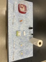
Safe handling of scalpels and proper slide preparation are critical for successful microscopy.
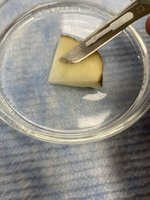
Cut away from your body and keep fingers clear of the blade.
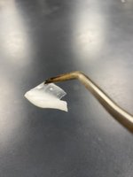
Use a cutting surface and make thin slivers for slides.
Prevent cross-contamination by using labeled tools.
Summary Table: Comparison of Cell Types
Feature | Prokaryotes | Eukaryotes |
|---|---|---|
Nucleus | No | Yes |
Organelles | No | Yes |
Cell Wall | Yes (most) | Plants: Yes; Animals: No |
Examples | Bacteria, Archaea | Plants, Animals, Fungi, Protists |
Size | Small (0.1–5 µm) | Larger (10–100 µm) |
Additional info:
Protists display a wide range of cellular structures and behaviors, making them important for understanding eukaryotic diversity.
Microscopy is a foundational technique in biology, allowing direct observation of cellular structures and functions.
