 뒤로
Cell–Cell Interactions: Structure, Communication, and Signaling
스터디 가이드 - 스마트 노트
 자료에 맞춘 맞춤형 노트, 핵심 정의, 예시, 맥락을 확장해 제공합니다.
Chapter 11: Cell–Cell Interactions
Overview and Roadmap
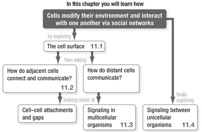
This chapter explores how cells modify their environment and interact with one another via social networks. The main topics include the cell surface, cell–cell attachments and gaps, signaling in multicellular organisms, and signaling between unicellular organisms.
Cell Walls in Prokaryotes
Prokaryotic cell wall structures differ between bacteria and archaea:
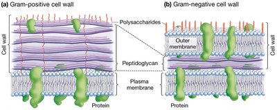
Bacteria: Cell walls consist primarily of polysaccharide peptidoglycan polymers connected by peptide bonds.
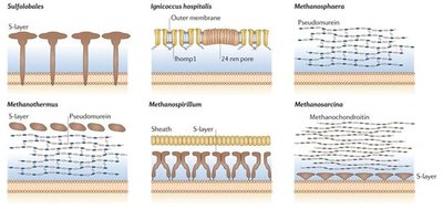
Archaea: Cell walls lack peptidoglycan and form a dense coat of proteins (S-layer).
 The Cell Surface
Structure and Function of the Plasma Membrane
The plasma membrane is a phospholipid bilayer embedded with proteins, which regulate transport and attach to cytoskeletal elements and extracellular structures. Most cells possess a protective layer or wall beyond the membrane, known as the extracellular matrix (ECM), which helps define cell shape, attach cells to one another, and acts as a first defense.
Integral proteins: Span the membrane and are involved in transport.
Peripheral proteins: Attach to the membrane surface and cytoskeleton.
Extracellular matrix (ECM): Defines cell shape, attaches cells, and provides defense.
Extracellular Layers in Eukaryotes
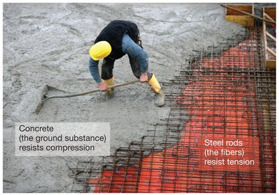
Eukaryotic extracellular layers share a fundamental organization, consisting of a fiber composite surrounded by a stiff ground substance. This structure resists tension and compression, providing mechanical support.
Fiber composite: Network of long filaments that resist tension.
Ground substance: Protects against compression.
Plant Cell Walls
Primary Cell Wall
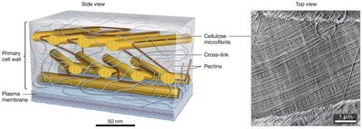
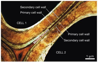
Most plant cells are surrounded by a cell wall. New plant cells secrete a fiber composite called the primary cell wall, which consists of cellulose microfibrils and pectin, forming a crisscrossed network that keeps the cell wall moist.
Secondary Cell Wall
Mature plant cells secrete a secondary cell wall between the plasma membrane and primary cell wall. Its structure correlates with cell function, containing waxes in leaf cells and lignin in wood-forming cells.
Extracellular Matrix in Animals
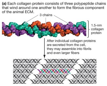
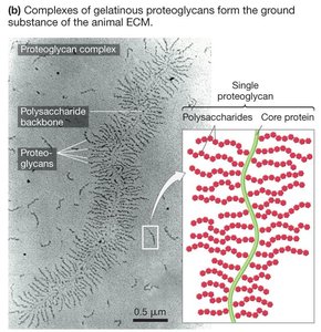
Most animal cells secrete a fiber composite called the extracellular matrix (ECM), which provides structural support. The fibrous component is mostly collagen, while the ground substance is made of proteoglycans, giving cartilage its rubber-like consistency.
Collagen: Triple helix structure forms fibrils and fibers.
Proteoglycans: Proteins attached to polysaccharides, forming the ground substance.
ECM Variation and Integrins
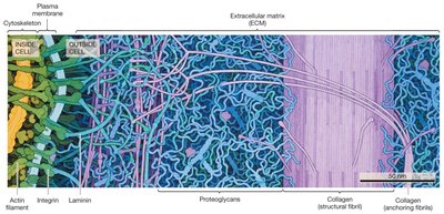
The amount and composition of ECM varies among tissues. Integrins are membrane proteins that bind to cross-linking proteins in the ECM, such as laminins, anchoring the cytoskeleton to the ECM and attaching the ECM to the plasma membrane.
How Do Adjacent Cells Connect and Communicate?
Cell–Cell Attachments in Multicellular Organisms
Direct physical connections between cells are essential for multicellularity, maintaining tissue structure and function. Cell–cell attachments are especially important in epithelia, tissues that line surfaces.
Indirect Cell–Cell Attachments in Plants
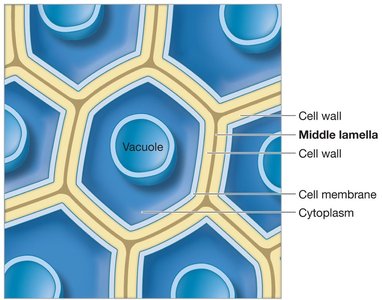
Plant cells are glued together by the middle lamella, a gelatinous layer continuous with adjacent cells' primary cell walls, comprised of pectins.
Tight Junctions
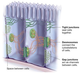
Tight junctions are cell–cell attachments composed of membrane proteins in adjacent animal cells, forming watertight seals. They are dynamic and variable, often found in epithelia, and can open or close in response to environmental changes.
Desmosomes
Desmosomes are strong cell–cell attachments common in animal epithelial and muscle cells. They comprise linking proteins and cytosolic anchoring proteins, with cytoskeletal intermediate filaments reinforcing the structure.
Gap Junctions
Gap junctions connect adjacent animal cells by forming channels that allow the flow of small molecules and ions, enabling rapid communication and coordination of activities.
Cell–Cell Signaling in Multicellular Organisms
Signaling Molecules and Reception
Cells not in physical contact can communicate via signaling molecules, such as neurotransmitters and hormones. Signal molecules deliver messages by binding to receptor proteins, which change shape and activity upon binding.
Signal receptors: Dynamic, can be blocked, and only cells with appropriate receptors respond.
Lipid-soluble signals: Diffuse across plasma membrane; receptors are in cytoplasm.
Lipid-insoluble signals: Recognized at cell surface; receptors are on plasma membrane.
Processing Lipid-Soluble and Lipid-Insoluble Signals
Lipid-soluble steroid hormones: Enter cytosol, form hormone–receptor complex, transported to nucleus, trigger changes in gene expression.
Lipid-insoluble hormones: Cannot diffuse across membrane; signal transduction converts extracellular signal to intracellular signal.
Signal Transduction Pathways
Signal transduction systems amplify and diversify messages:
G-protein-coupled receptors: Initiate production of second messengers, amplify signal.
Enzyme-linked receptors: Phosphorylate proteins inside target cell.
G-Protein-Coupled Receptor Pathway
Signaling molecule binds to membrane receptor.
G protein exchanges GDP for GTP; active subunit splits off.
Active G protein subunit activates nearby enzyme, catalyzing production of second messenger.
Second Messengers
Second messengers are small molecules that diffuse rapidly, amplify hormone signals, and activate protein kinases. Examples include calcium ions, cAMP, cGMP, DAG, and IP3.
Name | Type of Response |
|---|---|
Calcium ion (Ca2+) | Binds to calmodulin; activates proteins |
Cyclic adenosine monophosphate (cAMP) | Activates certain protein kinases |
Cyclic guanosine monophosphate (cGMP) | Opens ion channels; activates protein kinases |
Diacylglycerol (DAG) | Activates protein kinases |
Inositol trisphosphate (IP3) | Opens calcium channels |
Enzyme-Linked Receptor Pathway
Hormone binds to two RTK subunits, forming a dimer.
RTK is phosphorylated by ATP.
Proteins build a bridge between RTK and Ras protein, activating Ras.
Activated Ras phosphorylates protein kinase.
Triggers a phosphorylation cascade of protein kinases.
Signal Response and Deactivation
Signal transduction converts extracellular messages into intracellular responses, amplifying and diversifying the original message. Ultimate responses include changes in gene expression and protein activity. Cells have mechanisms for rapid signal deactivation, such as phosphatases removing phosphate groups.
Crosstalk Between Signaling Pathways
Crosstalk integrates diverse signals, allowing cells to respond in an integrated way. Pathways may inhibit or stimulate each other, and multiple steps provide points for crosstalk.
Signaling Between Unicellular Organisms
Quorum Sensing
Unicellular organisms communicate via signaling pathways that respond to population density. Bacteria release species-specific signaling molecules when their numbers reach a threshold, coordinating activities such as biofilm formation.
Quorum sensing: Allows unicellular organisms to coordinate activities.
G-protein coupled receptor: Causes free-living cells to aggregate (e.g., slime mold).
Summary Table: Connective Tissue of Animals
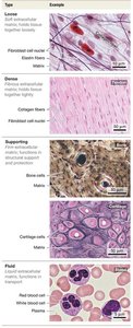
Connective tissues in animals are classified based on their extracellular matrix and function.
Type | Example |
|---|---|
Loose | Fibroblast cell nuclei, Collagen fibers |
Dense | Fibroblast cell nuclei, Collagen fibers |
Supporting | Bone matrix, Cartilage cells |
Fluid | Red blood cell, Plasma, White blood cell |
Morphological Diversity Among Bacteria
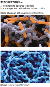
Bacteria exhibit extensive morphological diversity, including rods, chains of spheres, and spirals.
Types of Cell Walls in Bacteria and Archaea
Bacterial cell walls are classified as Gram-positive or Gram-negative based on their structure and staining properties. Archaea have diverse cell wall structures, often lacking peptidoglycan.
Fiber Composites: Tension and Compression
Fiber composites in extracellular layers resist tension and compression, similar to steel rods and concrete in construction.
Plant Cell Wall Structure
Cellulose microfibrils and pectin form the primary cell wall, providing structural support and moisture retention.
Secondary Cell Wall in Plants
The secondary cell wall is secreted by mature plant cells and varies in composition based on cell function.
Collagen and Proteoglycans in Animal ECM
Collagen forms the fibrous component, while proteoglycans form the ground substance of the animal ECM.
Integrins and ECM Connection
Integrins connect the cytoskeleton to the ECM, anchoring cells and providing structural integrity.
Cell–Cell Adhesion and Communication Structures
Tight junctions, desmosomes, and gap junctions are key structures in animal cell–cell adhesion and communication.
Middle Lamella in Plant Cells
The middle lamella connects adjacent plant cells, facilitating adhesion and communication.
Equations and Formulas
Phosphorylation Cascade:
G-Protein Activation:
Second Messenger Amplification:
Additional info: Academic context was added to clarify the function and structure of cell–cell interactions, signaling pathways, and extracellular matrices. All images included are directly relevant to the explanation of the adjacent paragraph.
