 뒤로
Chapter 5: The Working Cell – Membrane Structure, Function, and Cellular Energy
스터디 가이드 - 스마트 노트
 자료에 맞춘 맞춤형 노트, 핵심 정의, 예시, 맥락을 확장해 제공합니다.
Membrane Structure and Function
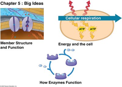
Fluid Mosaic Model of Cell Membranes
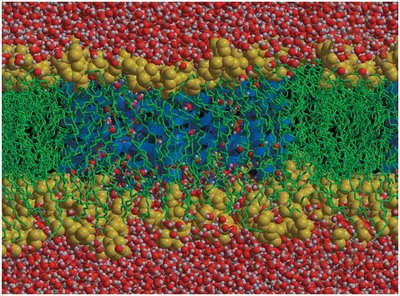
The fluid mosaic model describes the structure of cell membranes as a dynamic arrangement of diverse protein molecules suspended in a fluid phospholipid bilayer. This model explains how membranes are both flexible and selectively permeable, allowing cells to maintain homeostasis and interact with their environment.
Phospholipid bilayer: Composed of hydrophilic heads facing outward and hydrophobic tails facing inward, forming a semi-permeable barrier.
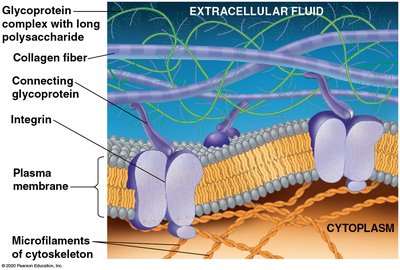
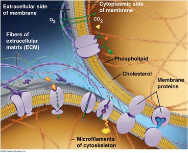
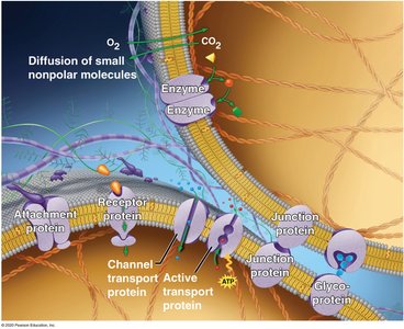
Membrane proteins: Embedded or attached to the bilayer, performing various functions such as transport, signaling, and structural support.
Selective permeability: The membrane allows certain substances to pass while restricting others.
Diverse Functions of Membrane Proteins
Membrane proteins are essential for the cell's interaction with its environment and internal regulation. They are classified based on their functions:
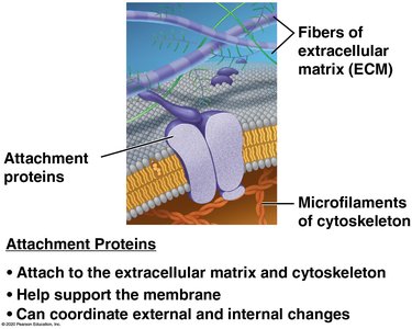
Attachment proteins: Connect the membrane to the extracellular matrix (ECM) and cytoskeleton, providing structural support and coordinating internal and external changes.
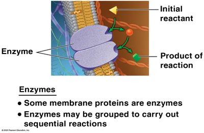
Enzymes: Catalyze sequential reactions, some grouped together for efficiency.
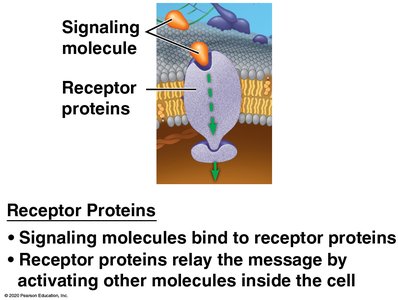
Receptor proteins: Bind signaling molecules and relay messages inside the cell, initiating cellular responses.
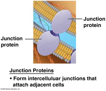
Junction proteins: Form intercellular junctions that attach adjacent cells, facilitating communication and cohesion.
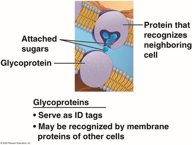
Glycoproteins: Serve as ID tags, recognized by other cells for cell-cell recognition.
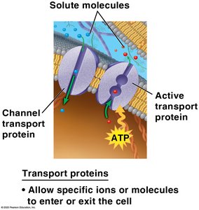
Transport proteins: Facilitate the movement of ions and molecules across the membrane, including channel and active transport proteins.
Transport Across Membranes
Passive Transport and Diffusion
Diffusion is the movement of particles from an area of higher concentration to lower concentration, driven by kinetic energy. When diffusion occurs across a cell membrane without energy input, it is called passive transport.
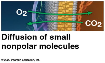
Simple diffusion: Small, nonpolar molecules (e.g., O2, CO2) pass directly through the membrane.
Facilitated diffusion: Polar or charged substances move across membranes with the help of transport proteins, down their concentration gradient.
Osmosis and Water Balance
Osmosis is the diffusion of water across a selectively permeable membrane. Water moves from areas of lower solute concentration to higher solute concentration until equilibrium is reached.
Tonicity: Describes the ability of a solution to cause a cell to gain or lose water.
Hypertonic solution: Higher solute concentration outside the cell; cell shrinks.
Hypotonic solution: Lower solute concentration outside the cell; cell swells.
Isotonic solution: Equal solute concentration; animal cells are normal, plant cells are flaccid.
Active Transport
Active transport requires energy (usually ATP) to move solutes against their concentration gradient. This process is essential for maintaining cellular concentrations of ions and other substances.
Transport proteins: Use ATP to move molecules from low to high concentration.
Bulk Transport: Exocytosis and Endocytosis
Cells use exocytosis to export large molecules and endocytosis to import them. Endocytosis includes phagocytosis (engulfing particles) and receptor-mediated endocytosis (specific uptake via receptors).
Exocytosis: Vesicles fuse with the membrane to release contents outside the cell.
Endocytosis: Membrane wraps around material to bring it into the cell.
Energy and the Cell
Types of Energy and Thermodynamics
Cells require energy to perform work. Energy exists in various forms:
Kinetic energy: Energy of motion.
Potential energy: Stored energy due to position or structure.
Chemical energy: Potential energy stored in chemical bonds.
Heat: Energy transferred due to temperature difference.
The first law of thermodynamics states that energy cannot be created or destroyed, only transformed. The second law states that energy transformations increase disorder (entropy), with some energy lost as heat.
Exergonic and Endergonic Reactions
Exergonic reactions release energy, while endergonic reactions require energy input. Cellular metabolism includes both types of reactions.
Exergonic: Cellular respiration, breakdown of molecules.
Endergonic: Synthesis of molecules, such as proteins.
ATP and Energy Coupling
ATP (adenosine triphosphate) is the cell's energy currency. It powers cellular work by transferring a phosphate group to other molecules, coupling exergonic and endergonic reactions.
ATP hydrolysis:
Energy coupling: Exergonic reactions provide energy for endergonic processes via ATP.
How Enzymes Function
Enzyme Structure and Catalysis
Enzymes are biological catalysts that speed up chemical reactions by lowering the activation energy required. They are not consumed in the reaction.
Active site: Region of the enzyme where the substrate binds.
Substrate: The reactant acted upon by the enzyme.
Catalytic cycle: Substrate binds, reaction occurs, product is released.
Enzyme Inhibition and Regulation
Enzyme activity can be regulated by inhibitors:
Competitive inhibitors: Block the active site, preventing substrate binding.
Noncompetitive inhibitors: Bind elsewhere, changing enzyme shape and reducing activity.
Feedback inhibition: End product of a pathway inhibits an earlier enzyme, regulating metabolism.
Enzyme Inhibitors: Drugs, Pesticides, and Poisons
Many drugs, pesticides, and poisons act as enzyme inhibitors, affecting cellular processes. The reversibility of inhibition depends on the nature of the inhibitor and its binding to the enzyme.
Tables: Enzyme Activity
Reaction Rate and Enzyme Concentration
This table shows how increasing enzyme concentration affects the reaction rate when substrate concentration is constant.
Lactose concentration | Enzyme concentration | Reaction rate |
|---|---|---|
10% | 0% | 0 |
10% | 1% | 25 |
10% | 2% | 50 |
10% | 4% | 100 |
10% | 8% | 200 |
Reaction Rate and Substrate Concentration
This table shows how increasing substrate concentration affects the reaction rate when enzyme concentration is constant.
Lactose concentration | Enzyme concentration | Reaction rate |
|---|---|---|
0% | 2% | 0 |
5% | 2% | 25 |
10% | 2% | 50 |
20% | 2% | 65 |
30% | 2% | 65 |
Interpretation: Reaction rate increases with enzyme concentration (Table A) and with substrate concentration up to a point, after which it plateaus (Table B), indicating enzyme saturation.
