 뒤로
Circulation and Gas Exchange: Structure, Function, and Regulation in Animals
스터디 가이드 - 스마트 노트
 자료에 맞춘 맞춤형 노트, 핵심 정의, 예시, 맥락을 확장해 제공합니다.
Circulation and Gas Exchange
Overview: Trading with the Environment
All organisms must exchange materials with their environment, a process that ultimately occurs at the cellular level. In unicellular organisms, exchanges happen directly with the environment, but multicellular organisms require specialized systems to facilitate these exchanges.
Direct exchange is possible for unicellular organisms.
Specialized exchange systems (e.g., gills in fish) are necessary for multicellular organisms.
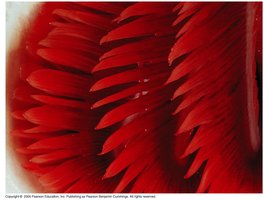
Example: Salmon gills are highly adapted for efficient gas exchange.
Concept 42.1: Circulatory Systems Link Exchange Surfaces with Cells Throughout the Body
Transport systems connect organs of exchange with body cells. Most complex animals have internal transport systems that circulate fluid, ensuring efficient distribution of substances.
Diffusion is inadequate for large organisms; circulatory systems reduce diffusion distances.
Closed circulatory system (cardiovascular system) in vertebrates consists of blood vessels and a heart with chambers.
Arteries carry blood away from the heart; veins return blood to the heart; capillaries are sites of exchange.
Blood vessel number correlates with organ metabolic requirements.
Survey of Vertebrate Circulation
Vertebrates exhibit diverse heart and circulatory system structures, reflecting evolutionary adaptations.
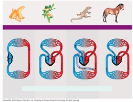
Fish: Two-chambered heart (one atrium, one ventricle); blood flows through gills for gas exchange, then systemic circulation.
Amphibians: Three-chambered heart (two atria, one ventricle); ventricle pumps blood to pulmocutaneous (lungs/skin) and systemic circuits.
Reptiles (except birds): Three-chambered heart with partial septum; double circulation (pulmonary and systemic circuits).
Mammals and Birds: Four-chambered heart; complete separation of oxygen-rich and oxygen-poor blood; essential for endothermy.
Concept 42.2: Coordinated Cycles of Heart Contraction Drive Double Circulation in Mammals
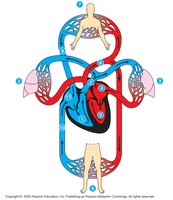
The mammalian circulatory system is a model for double circulation, ensuring efficient oxygen delivery and waste removal.
Heart valves ensure one-way blood flow.
Pathway: Right ventricle → lungs (O2 uptake, CO2 release) → left atrium → left ventricle → body tissues → right atrium.
The Mammalian Heart: A Closer Look
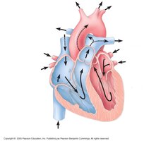
The heart consists mostly of cardiac muscle and operates in a rhythmic cycle.
Atria contract to fill ventricles.
Ventricles pump blood to organs.
Valves prevent backflow (atrioventricular and semilunar valves).
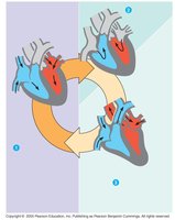
The Cardiac Cycle
The heart contracts and relaxes in a rhythmic cycle:
Systole: Contraction/pumping phase.
Diastole: Relaxation/filling phase.
Heart Rate and Cardiac Output
Heart rate (pulse): Beats per minute.
Cardiac output: Volume of blood pumped per minute ().
Typical value: 5.25 L/min.
Valves and Heart Sounds
Atrioventricular (AV) valves: Between atria and ventricles; prevent backflow into atria.
Semilunar valves: At exits of heart; prevent backflow into ventricles.
Heart sounds: "Lub" (AV valves close), "Dub" (semilunar valves close).
Maintaining the Heart’s Rhythmic Beat
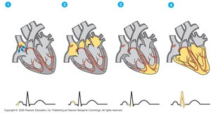
Self-excitable cardiac muscle cells contract without nervous input.
Sinoatrial (SA) node: Pacemaker; sets contraction rate.
Atrioventricular (AV) node: Delays signal, then sends to Purkinje fibers for ventricular contraction.
Electrocardiogram (ECG): Records electrical impulses during cardiac cycle.
Regulation of Pacemaker Activity
Influenced by nerves, hormones, temperature, and exercise.
Increase in temperature or exercise raises heart rate.
Concept 42.3: Physical Principles Govern Blood Circulation
Blood vessel structure and function are governed by physical principles similar to plumbing systems.
Blood Vessel Structure
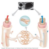
Three layers: connective tissue, smooth muscle, endothelium.
Capillaries: Only endothelium and basement membrane; facilitate exchange.
Arteries: Thick walls for high pressure.
Veins: Thinner walls; rely on muscle action and valves for blood return.
Blood Flow in Veins
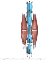
Skeletal muscle contraction aids venous return.
Valves prevent backflow.
Blood Flow Velocity
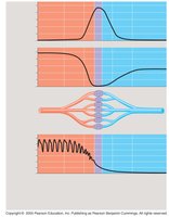
Fluid flows faster in narrow segments; slowest in capillaries due to high resistance and large cross-sectional area.
Blood Pressure
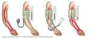
Systolic pressure: Highest pressure during ventricular contraction.
Diastolic pressure: Lower pressure during relaxation.
Blood pressure depends on cardiac output and peripheral resistance.
Capillary Function
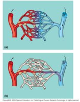
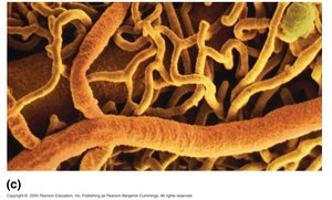
Blood supply to capillary beds is regulated by smooth muscle contraction and precapillary sphincters.
Exchange of substances occurs across thin endothelial walls.
Fluid movement is driven by differences in blood pressure and osmotic pressure.
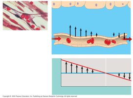
Fluid Return by the Lymphatic System
Lymphatic system returns fluid from capillary beds to circulation.
Aids in body defense; lymph nodes filter lymph.
Concept 42.4: Blood is a Connective Tissue with Cells Suspended in Plasma
Blood Composition and Function
Plasma: 90% water; contains electrolytes, plasma proteins, nutrients, waste, gases, hormones.
Cellular elements: Erythrocytes (red blood cells), leukocytes (white blood cells), platelets.
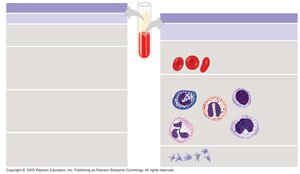
Erythrocytes
Most numerous; transport oxygen via hemoglobin.
Lack nuclei and mitochondria; biconcave shape increases surface area.
Leukocytes
Five major types: monocytes, neutrophils, basophils, eosinophils, lymphocytes.
Defense and immunity; lymphocytes produce antibodies.
Platelets
Fragments of cells; involved in blood clotting.
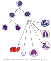
Stem Cells and Replacement of Cellular Elements
All blood cells develop from pluripotent stem cells in bone marrow.
Erythropoietin (EPO) stimulates erythrocyte production in response to low oxygen.
Leukemia: Overproduction of leukocytes impairs red blood cell formation.
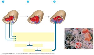
Blood Clotting
Damage to vessel initiates clotting cascade: fibrinogen → fibrin.
Platelets form a plug; clot reinforces seal.
Hemophilia: Defects in clotting process.
Cardiovascular Disease
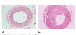
Includes atherosclerosis, hypertension, heart attack, stroke.
Atherosclerosis: Cholesterol buildup in arteries; LDL (bad), HDL (good).
Hypertension increases risk of heart attack and stroke.
Concept 42.5: Gas Exchange Occurs Across Specialized Respiratory Surfaces
Gas exchange supplies oxygen for cellular respiration and removes carbon dioxide. Large, moist respiratory surfaces are required for efficient diffusion.
Size of respiratory surface correlates with metabolic needs.
Lungs
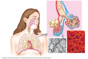
Internal lungs in terrestrial vertebrates; size and complexity reflect metabolic rate.
Branching ducts convey air to alveoli, the site of gas exchange.
Concept 42.6: Breathing Ventilates the Lungs
How a Mammal Breathes
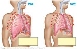
Negative pressure breathing: Rib muscles and diaphragm contract, increasing lung volume and drawing air in.
Tidal volume: Air inhaled/exhaled per breath (500 mL).
Vital capacity: Maximum tidal volume during forced breathing.
Residual volume: Air remaining after forceful exhale.
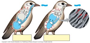
How a Bird Breathes
Birds have air sacs that keep air flowing in one direction through lungs.
Parabronchi are sites of gas exchange; efficient at high altitudes.
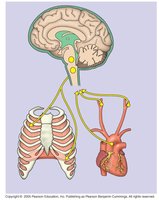
Control of Breathing in Humans
Breathing control centers in medulla oblongata and pons.
Medulla responds to pH changes in cerebrospinal fluid; sensors in aorta and carotid arteries monitor O2 and CO2 levels.
Concept 42.7: Respiratory Pigments Bind and Transport Gases
The Role of Partial Pressure Gradients
Gases diffuse down partial pressure gradients.
Partial pressure of O2 in air:
O2 and CO2 move from high to low partial pressure regions.
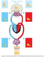
Respiratory Pigments
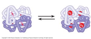
Hemoglobin is the main respiratory pigment in vertebrates; increases O2 carrying capacity.
Each hemoglobin molecule binds four O2 molecules via iron atoms.
Binding and release of O2 are cooperative among hemoglobin subunits.
Oxygen Dissociation and the Bohr Effect
O2 unloading increases in tissues with high metabolism or lower pH (Bohr shift).
Carbon Dioxide Transport
Hemoglobin also transports CO2 and buffers blood.
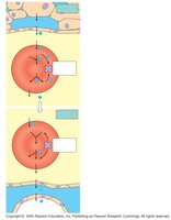
CO2 is converted to bicarbonate () in erythrocytes and released in lungs.
Additional info: These notes expand on the original content with definitions, examples, and academic context to provide a comprehensive study guide for Chapter 42: Circulation and Gas Exchange in General Biology.
