 뒤로
DNA Structure and Replication: Foundations of Genetic Inheritance
스터디 가이드 - 스마트 노트
 자료에 맞춘 맞춤형 노트, 핵심 정의, 예시, 맥락을 확장해 제공합니다.
DNA and Chromosomes
Organization of Genetic Material
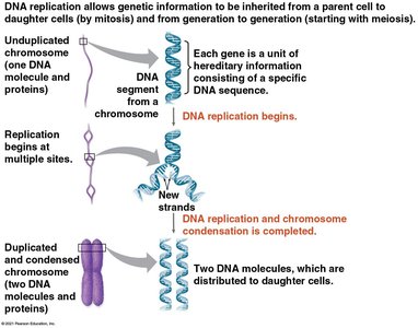
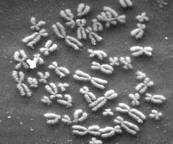
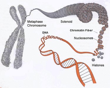
DNA is the hereditary material in almost all living organisms, organized into structures called chromosomes. Each chromosome consists of a single, long DNA molecule wrapped around proteins called histones, forming a compact structure that fits within the cell nucleus.
Chromatin: The combination of DNA and proteins (mainly histones) that make up chromosomes.
Metaphase Chromosome: The most condensed form of chromatin, visible during cell division.
Gene: A segment of DNA that encodes a specific protein or functional RNA.
Structure of DNA
Nucleotide Composition and Base Pairing
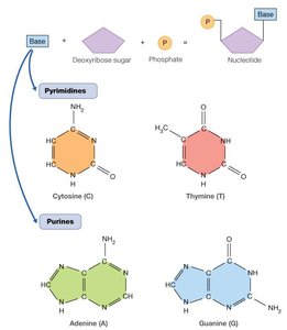
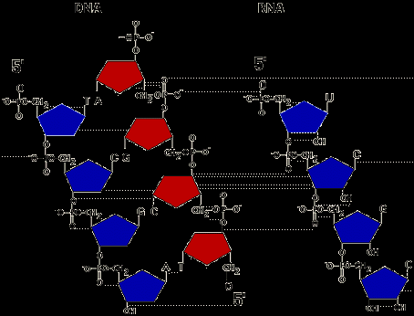
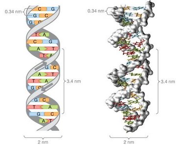
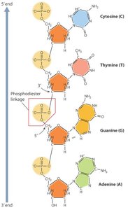
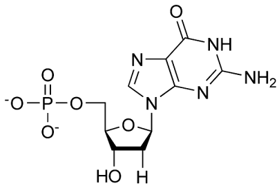
DNA is a polymer made of repeating units called nucleotides. Each nucleotide consists of a deoxyribose sugar, a phosphate group, and a nitrogenous base. The four types of nitrogenous bases in DNA are adenine (A), thymine (T), cytosine (C), and guanine (G).
Pyrimidines: Cytosine (C) and Thymine (T) – single-ring structures.
Purines: Adenine (A) and Guanine (G) – double-ring structures.
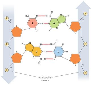
Base Pairing: A pairs with T (via 2 hydrogen bonds), and G pairs with C (via 3 hydrogen bonds), according to Chargaff’s rules.
DNA Backbone and Directionality
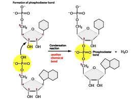
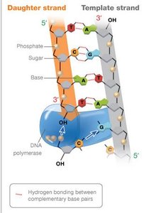
The backbone of DNA is formed by alternating sugar and phosphate groups, connected by phosphodiester bonds. DNA strands have directionality, with a 5’ (phosphate) end and a 3’ (hydroxyl) end. During polymerization, the 5’ phosphate of one nucleotide joins the 3’ hydroxyl of another.
Phosphodiester Bond: The covalent bond linking nucleotides in a DNA strand.
Antiparallel Strands: The two DNA strands run in opposite directions (5’→3’ and 3’→5’).
Discovery of DNA as Genetic Material
Key Experiments
Several classic experiments established DNA as the genetic material:
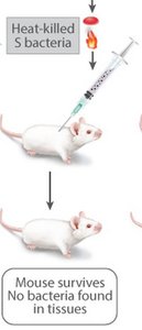
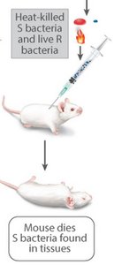
Griffith’s Experiment (1928): Demonstrated transformation in bacteria, showing that genetic material could be transferred between cells.

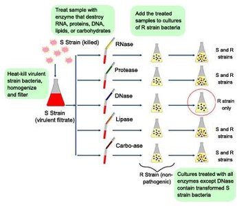
Avery, MacLeod, and McCarty (1944): Identified DNA as the transforming principle by selectively destroying biomolecules and observing transformation.
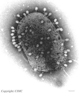
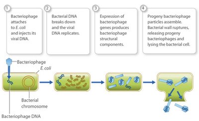
Hershey-Chase Experiment (1952): Used bacteriophages labeled with radioactive isotopes to show that DNA, not protein, enters bacterial cells and directs viral replication.
The Double Helix Model
Watson and Crick’s Discovery
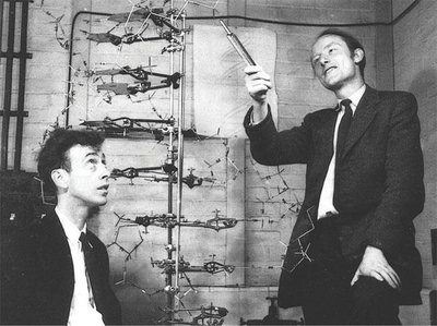
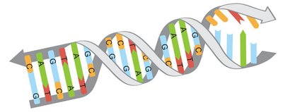
James Watson and Francis Crick, with contributions from Rosalind Franklin and Maurice Wilkins, proposed the double helix structure of DNA in 1953. The model explained how genetic information is stored and replicated.
Antiparallel Strands: The two DNA strands run in opposite directions.
Hydrogen Bonding: Base pairs are held together by hydrogen bonds.
Major and Minor Grooves: The double helix has grooves that allow protein binding.
DNA Replication
Models of Replication
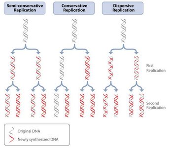
Three models were proposed for DNA replication: conservative, semi-conservative, and dispersive. The semi-conservative model, supported by the Meselson-Stahl experiment, is correct: each new DNA molecule consists of one old (parental) strand and one newly synthesized strand.
Semi-conservative: Each daughter DNA has one parental and one new strand.
Conservative: Parental DNA remains intact; new DNA is entirely new.
Dispersive: Each strand is a mix of old and new DNA.
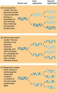
Mechanism of DNA Replication
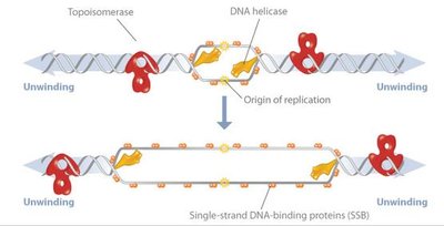
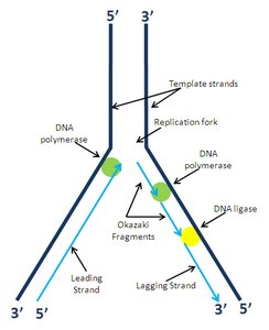
DNA replication is a complex, enzyme-driven process that ensures accurate copying of genetic material. It begins at specific sites called origins of replication and proceeds bidirectionally, forming replication forks.
Initiation: DNA helicase unwinds the double helix, and single-stranded binding proteins stabilize the unwound strands.
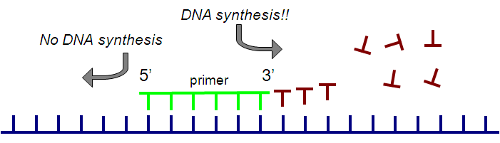
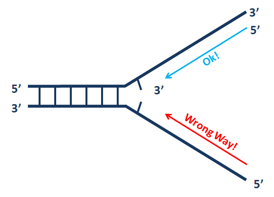
Elongation: DNA polymerase synthesizes new DNA by adding nucleotides to the 3’ end of a primer.
Leading Strand: Synthesized continuously in the 5’→3’ direction.
Lagging Strand: Synthesized discontinuously as Okazaki fragments, later joined by DNA ligase.
Primase: Synthesizes short RNA primers to provide a starting point for DNA polymerase.
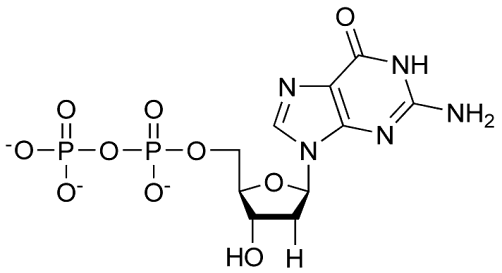
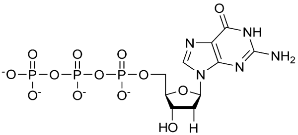
Energy Source: Nucleotides are added as triphosphates (e.g., dATP), and the release of pyrophosphate provides energy for bond formation.
Enzymes and Proteins in DNA Replication
Enzyme/Protein | Function | Location |
|---|---|---|
Helicase | Unwinds the DNA double helix | Replication fork |
DNA Polymerase III | Main enzyme for DNA synthesis | Replication fork |
DNA Polymerase I | Removes RNA primers and fills in gaps | Replication fork |
Primase | Synthesizes RNA primers | Replication fork |
Ligase | Joins Okazaki fragments | Lagging strand |
Telomerase | Extends telomeres in eukaryotes | Chromosome ends |
Proofreading and DNA Repair
DNA polymerases have proofreading ability, correcting most errors during replication. Additional repair mechanisms, such as mismatch repair and excision repair, further ensure fidelity. Unrepaired errors can result in mutations.
Replication of Chromosome Ends: Telomeres and Telomerase
Linear chromosomes in eukaryotes face the end-replication problem, where the very ends (telomeres) cannot be fully replicated by standard DNA polymerases. Telomerase extends telomeres, preventing loss of genetic information in germ cells and some stem cells.
In Vitro DNA Replication: Polymerase Chain Reaction (PCR)
PCR vs. Cellular DNA Replication
PCR is a laboratory technique used to amplify specific DNA sequences. It requires template DNA, DNA primers, nucleotides, a heat-stable DNA polymerase, and a buffer. The process involves repeated cycles of denaturation, annealing, and extension.
Similarities: Both require primers, nucleotides, DNA template, and polymerase; both synthesize DNA in the 5’→3’ direction.
Differences: PCR uses DNA primers and heat to separate strands, while cellular replication uses RNA primers and helicase; PCR amplifies specific regions, while cellular replication copies entire chromosomes.
Summary Table: Key Enzymes in DNA Replication
Enzyme | Function | Found In |
|---|---|---|
Helicase | Unwinds DNA double helix | All cells |
Ligase | Seals nicks, joins Okazaki fragments | All cells |
DNA Polymerase I | Removes RNA primers, fills gaps | Prokaryotes |
DNA Polymerase III | Main DNA synthesis enzyme | Prokaryotes |
Primase | Synthesizes RNA primers | All cells |
Telomerase | Extends telomeres | Eukaryotes (germ/stem/cancer cells) |
Key Equations and Concepts
Phosphodiester Bond Formation:
Base Pairing Rule:
Directionality: DNA synthesis always proceeds 5’→3’.
Additional info: The notes above integrate foundational knowledge from Chapters 5 and 16 of a typical General Biology curriculum, covering DNA structure, function, and replication, as well as experimental evidence and modern applications such as PCR.
