 뒤로
뒤로General Biology Study Notes: Scientific Method, Chemistry of Life, Macromolecules, and Cell Structure
스터디 가이드 - 스마트 노트
 자료에 맞춘 맞춤형 노트, 핵심 정의, 예시, 맥락을 확장해 제공합니다.
Scientific Method and Laboratory Safety
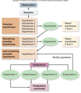
Overview of the Scientific Method
The scientific method is a systematic approach used by scientists to investigate natural phenomena, acquire new knowledge, or correct and integrate previous knowledge. It involves making observations, forming hypotheses, conducting experiments, collecting data, and drawing conclusions.
Observation: Gathering information about a phenomenon or problem.
Question: Formulating a question based on observations.
Hypothesis: Proposing a testable explanation or prediction.
Experiment: Designing and performing tests to evaluate the hypothesis.
Data: Collecting and analyzing results from experiments.
Conclusion: Interpreting data to accept, reject, or modify the hypothesis.

Scientific investigations often involve multiple hypotheses and iterative testing, leading to refined predictions and further experimentation.
Laboratory Safety and PPE
Personal Protective Equipment (PPE) is essential for safety in the laboratory. Proper attire includes lab coats, safety goggles or glasses, and gloves. PPE protects against chemical spills, biological samples, and sharp objects, reducing the risk of injury or contamination.
Lab coat: Protects skin and clothing from spills.
Safety goggles/glasses: Shields eyes from splashes and debris.
Gloves: Prevents direct contact with hazardous substances.

Proper use of PPE is a professional habit and a requirement for participation in laboratory activities.
Laboratory Notebook Structure
A laboratory notebook is a critical tool for documenting experiments. It should include:
Title: Describes the experiment.
Introduction: States objectives, purpose, and background.
Hypothesis: A testable prediction based on observations.
Procedures: Step-by-step methods and protocols.
Results: Data collected, often in tables or graphs.
Discussion: Interpretation of results, conclusions, and answers to guiding questions.
Chemistry in Biology: Measurement and pH
Metric System and Units of Measurement
The metric system is the universal standard for scientific measurement. It is based on units of ten, making conversions straightforward. Key units include:
Measure | Unit | Conversion |
|---|---|---|
Length | meter (m) | 1 m = 100 cm = 1000 mm |
Mass | gram (g) | 1 g = 1000 mg |
Volume | liter (L) | 1 L = 1000 mL |
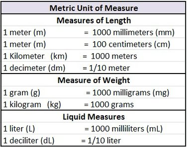
For water, 1 g = 1 cm3 = 1 mL. For other substances, density must be considered for conversions.
pH: Acids, Bases, and Buffers
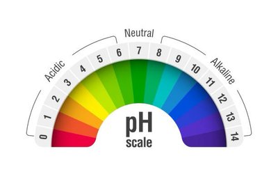
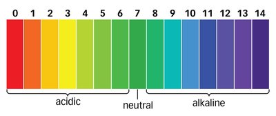
The pH scale measures the concentration of hydrogen ions (H+) in a solution, indicating its acidity or alkalinity. The scale ranges from 0 (very acidic) to 14 (very basic), with 7 being neutral.
Acids: pH < 7, high H+ concentration.
Bases (Alkaline): pH > 7, low H+ concentration.
Neutral: pH = 7 (pure water).
Organisms function best near neutral pH, but some specialize in acidic (acidophiles) or basic (alkalinophiles) environments. Buffers are systems of weak acids or bases that resist changes in pH, maintaining homeostasis in biological systems.
Table: pH Classification of Common Substances
Substance | Acid/Base/Neutral | Estimated pH |
|---|---|---|
Tomato Juice | Acid | ~4 |
Cola | Acid | ~2-3 |
Lime Juice | Acid | ~2 |
Vinegar | Acid | ~3 |
Ammonia | Base | ~11 |
Baking Soda (NaHCO3) | Base | ~9 |
Salicylic Acid (aspirin) | Acid | ~3 |
Milk | Acid | ~6.5 |
NaCl (salt) | Neutral | ~7 |
Urea | Neutral | ~7 |
Distilled Water | Neutral | 7 |
*Additional info: Estimated pH values are based on typical laboratory measurements.
Macromolecules: Structure and Function
Overview of Biological Macromolecules
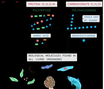
All living organisms are composed of four major classes of macromolecules: carbohydrates, lipids, proteins, and nucleic acids. These molecules are primarily carbon-based and are essential for structure, function, and information storage in cells.
Carbohydrates: Main energy source and structural support (e.g., cellulose, chitin).
Lipids: Long-term energy storage, cell membrane structure, hormones.
Proteins: Structure, enzymes, transport, signaling.
Nucleic Acids: Store and transmit genetic information (DNA, RNA).
Monomers and Polymers
Macromolecules are polymers built from smaller units called monomers:
Carbohydrates: Monosaccharides
Lipids: Glycerol + Fatty acids
Proteins: Amino acids
Nucleic Acids: Nucleotides

Carbohydrates
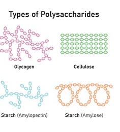
Carbohydrates are composed of carbon, hydrogen, and oxygen. They serve as energy sources and structural components. Polysaccharides such as starch, glycogen, and cellulose are polymers of glucose.
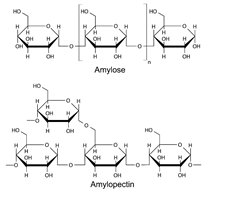
Starch: Energy storage in plants; can be amylose (straight-chain) or amylopectin (branched).
Glycogen: Energy storage in animals; highly branched.
Cellulose: Structural component in plant cell walls; composed of beta-D-glucose, indigestible by mammals.
Chitin is a nitrogen-containing polysaccharide found in arthropod exoskeletons and fungal cell walls.
Proteins
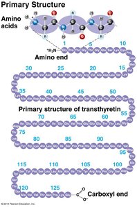
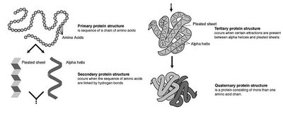
Proteins are polymers of amino acids, which contain a central (alpha) carbon, an amino group, a carboxyl group, a hydrogen atom, and a variable R group. The sequence and properties of amino acids determine protein structure and function.

Primary structure: Linear sequence of amino acids.
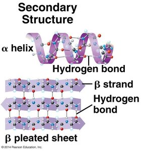
Secondary structure: Alpha helices and beta sheets formed by hydrogen bonding.
Tertiary structure: 3D folding due to interactions among R groups (hydrogen bonds, ionic bonds, hydrophobic interactions, disulfide bridges).
Quaternary structure: Association of multiple polypeptide chains.
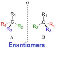
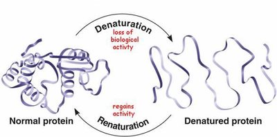
Proteins can be denatured (lose their structure and function) by changes in temperature or pH, but some can renature if conditions return to normal.
Lipids
Lipids are hydrophobic molecules, including fats, oils, phospholipids, waxes, and steroids. They are primarily composed of carbon and hydrogen, with some oxygen.
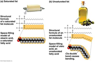
Triglycerides: Glycerol + 3 fatty acids; energy storage. Saturated fats (no double bonds) are solid at room temperature; unsaturated fats (one or more double bonds) are liquid.
Phospholipids: Glycerol + 2 fatty acids + phosphate group; major component of cell membranes, amphipathic (hydrophilic head, hydrophobic tails).
Steroids: Four-ring structure; includes cholesterol, hormones like estrogen and testosterone.
Cell Structure and Microscopy
Cell Theory and Types of Cells
The cell is the fundamental unit of life. According to cell theory, all living organisms are composed of cells, and all cells arise from pre-existing cells. Cells can be unicellular or multicellular.
Prokaryotic cells: Lack a nucleus and membrane-bound organelles; DNA is in the nucleoid region. Domains: Bacteria and Archaea.
Eukaryotic cells: Have a nucleus and membrane-bound organelles; found in plants, animals, fungi, and protists.
Microscopy
Microscopes are essential tools for studying cells and their structures. The compound light microscope uses visible light and multiple lenses to magnify specimens up to 1000x. Electron microscopes provide much higher magnification and resolution.
Ocular lens: Eyepiece, usually 10x magnification.
Objective lenses: Varying magnifications (4x, 10x, 40x, 100x).
Stage: Platform for holding slides.
Condenser and iris diaphragm: Focus and control light.
Coarse and fine focus knobs: Adjust focus.
Proper use and care of the microscope are essential for accurate observation and safety.
Cellular Organelles and Their Functions
Eukaryotic cells contain specialized organelles, each with distinct functions:
Nucleus: Contains DNA, site of replication and transcription.
Ribosomes: Protein synthesis; found free in cytosol or bound to rough ER.
Endoplasmic Reticulum (ER): Rough ER synthesizes proteins; smooth ER synthesizes lipids and detoxifies chemicals.
Golgi apparatus: Modifies, sorts, and packages proteins and lipids for transport.
Lysosomes: Contain hydrolytic enzymes for digestion of macromolecules.
Vacuoles: Storage and maintenance of cell turgor (central vacuole in plants).
Mitochondria: Site of cellular respiration and ATP production; double-membraned, contain their own DNA.
Chloroplasts: Site of photosynthesis in plants; double-membraned, contain chlorophyll and their own DNA.
Peroxisomes: Break down fatty acids and detoxify harmful substances.
Cytoskeleton: Provides structural support, cell shape, and movement.
Membrane Structure and Transport
Fluid Mosaic Model
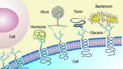
The fluid mosaic model describes the structure of the cell membrane as a dynamic, flexible bilayer of phospholipids with embedded proteins, cholesterol, and carbohydrates. The amphipathic nature of phospholipids (hydrophilic heads, hydrophobic tails) allows for selective permeability.
Integral proteins: Span the membrane, involved in transport and signaling.
Peripheral proteins: Attached to membrane surface, involved in signaling and structure.
Cholesterol: Modulates membrane fluidity and stability.
Membrane Transport Mechanisms
Passive transport: Movement of substances down their concentration gradient without energy input (e.g., diffusion, osmosis, facilitated diffusion).
Active transport: Movement of substances against their concentration gradient, requiring energy (ATP), e.g., sodium-potassium pump.
Bulk transport: Endocytosis (phagocytosis, pinocytosis, receptor-mediated) and exocytosis for large molecules or particles.
Homeostasis is maintained by selective permeability and regulated transport of ions, nutrients, and waste products.
Enzymes and Metabolism
Enzyme Structure and Function
Enzymes are biological catalysts, usually proteins, that speed up chemical reactions by lowering the activation energy required. Each enzyme is specific to its substrate, binding at the active site to form an enzyme-substrate complex.
Active site: Region on the enzyme where the substrate binds.
Cofactors: Non-protein molecules (organic or inorganic) required for enzyme activity (e.g., vitamins, metal ions).
Enzyme activity: Influenced by temperature, pH, substrate concentration, and presence of inhibitors or activators.
Enzymes can be inhibited competitively (inhibitor binds active site) or noncompetitively (allosteric inhibitor binds elsewhere, changing enzyme shape).
Enzyme Kinetics and Regulation
Reaction rate: Increases with substrate concentration until saturation is reached.
Temperature and pH: Each enzyme has optimal conditions; deviations can denature the enzyme and reduce activity.
Allosteric regulation: Binding of molecules at sites other than the active site can enhance or inhibit enzyme function.
Spectrophotometry and Beer-Lambert Law
Spectrophotometry measures the absorbance of light by a solution to determine the concentration of solutes. The Beer-Lambert Law states:
A: Absorbance
\varepsilon: Molar extinction coefficient (constant for a given substance and wavelength)
c: Concentration of the solute
l: Path length of light through the sample (usually 1 cm)
Absorbance is directly proportional to concentration, allowing quantification of reaction rates in enzyme assays.
