 뒤로
뒤로Lipids, Membranes, and the First Cells: Structure, Function, and Transport
스터디 가이드 - 스마트 노트
 자료에 맞춘 맞춤형 노트, 핵심 정의, 예시, 맥락을 확장해 제공합니다.
Lipids, Membranes, and the First Cells
Plasma Membrane: Structure and Function
The plasma membrane (or cell membrane) is a fundamental structure that separates living cells from their external environment. It acts as a selective barrier, allowing entry of essential materials and preventing the entry of harmful substances. The membrane also facilitates chemical reactions necessary for life by sequestering appropriate chemicals.
Lipid Structure and Function
Lipids are carbon-containing compounds that are insoluble in water due to their high proportion of nonpolar bonds. Hydrocarbons are nonpolar molecules containing only carbon and hydrogen, making them hydrophobic.
Isoprenoids: Hydrocarbon chains functioning as pigments, scents, vitamins, and hormone precursors; building blocks for complex lipids.
Fatty acids: Hydrocarbon chains bonded to a carboxyl functional group, containing 14–20 carbon atoms; can be saturated or unsaturated.
Bond Saturation and Hydrocarbon Structure
Saturation affects the physical state and structure of lipids:
Saturated fatty acids: Only single bonds between carbons; maximum hydrogen atoms; solid at room temperature.
Unsaturated fatty acids: One or more double bonds; forms a "kink" in the chain; liquid at room temperature; polyunsaturated chains have many double bonds.
Example: Foods with unsaturated lipids are considered healthier than those with saturated lipids.


Types of Lipids Found in Cells
Lipids do not possess a shared chemical structure; their hydrocarbon skeletons can be assembled in various ways. The three most important types of lipids in cells are:
Steroids: Bulky, four-ring structure; differ by functional groups attached to rings. Examples include hormones (estrogen, testosterone) and cholesterol.
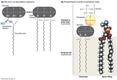
Fats: Three fatty acids linked to glycerol (triacylglycerols or triglycerides); primary role is energy storage.
Phospholipids: Glycerol linked to a phosphate group and two hydrocarbon chains; primary role is forming cell membranes.



How Membrane Lipids Interact with Water
Lipids serve multiple functions: storing chemical energy, acting as pigments, serving as signals, forming waterproof coatings, and acting as vitamins. Lipids contain both hydrophobic and hydrophilic regions:
Hydrophilic head: Contains glycerol, negatively charged phosphate group, and a charged or polar group.
Hydrophobic tail: Nonpolar, water molecules cannot form hydrogen bonds with the tail.
Phospholipids are amphipathic (having both hydrophilic and hydrophobic regions), which is crucial for plasma membrane formation.
Phospholipid Bilayers
Formation and Structure
Amphipathic lipids do not dissolve in water. Their hydrophilic heads interact with water, while hydrophobic tails do not. Amphipathic lipids form:
Micelles: Tiny spherical aggregates formed from free fatty acids.
Lipid bilayers: Paired sheets formed when lipid molecules align.

Phospholipid bilayers form spontaneously in water, decreasing entropy at the level of lipid organization due to aggregation of amphipathic lipids.
Artificial Membranes as Experimental Systems
Vesicles (liposomes) can be formed from phospholipids in the lab, serving as artificial membrane-bound vesicles. Planar bilayers are constructed across holes in glass or plastic walls to measure permeability.


Selective Permeability of Lipid Bilayers
Phospholipid bilayers exhibit selective permeability:
Small or nonpolar molecules move across quickly.
Charged or large polar substances cross slowly, if at all.
Examples: Oxygen (small, nonpolar) moves quickly; glucose (large, polar) moves slowly.

Factors Affecting Membrane Permeability
Length and saturation of hydrocarbon tails
Presence of cholesterol molecules
Unsaturated tails with double bonds create kinks, weakening the barrier to solutes. Saturated tails have stronger hydrophobic interactions, making membranes denser and less permeable.

Cholesterol increases the density of the hydrophobic section, reducing membrane permeability by forcing phospholipid tails closer together.
Temperature Effects
Membrane fluidity decreases as temperature drops, causing molecules to move more slowly and hydrophobic tails to pack tightly, resulting in decreased permeability.
Movement Across Lipid Bilayers: Diffusion and Osmosis
Diffusion
Diffusion is the spontaneous movement of molecules and ions due to their thermal energy, moving from regions of high concentration to low concentration. Diffusion increases entropy and leads to equilibrium, where molecules are randomly distributed. Passive transport occurs when substances diffuse across membranes without external energy.
Osmosis
Osmosis is a special case of diffusion involving water across selectively permeable membranes. Water moves from regions of low solute concentration to high solute concentration, diluting the higher concentration and equalizing concentrations on both sides.
Hypertonic: Higher solute concentration outside cell; water moves out, cell shrinks.
Hypotonic: Lower solute concentration outside cell; water moves in, cell swells.
Isotonic: Equal solute concentration; no net water movement, cell size remains constant.
Membranes and Chemical Evolution
The first lipid bilayers likely provided containers for replicating molecules such as RNA. Protocells are simple vesicle-like structures that harbor nucleic acids and may have been intermediates in the evolution of cells.
Proteins Alter Membrane Structure and Function
Membrane Proteins
Plasma membranes contain as much protein as phospholipids. Proteins can insert into membranes due to their amphipathic nature, forming openings and passageways across the lipid bilayer.
Fluid-Mosaic Model
The fluid-mosaic model describes the membrane as a dynamic mosaic of phospholipids and proteins, with some proteins spanning the membrane. This model replaced the earlier "sandwich" model after freeze-fracture electron microscopy revealed the true nature of membrane proteins.
Integral (transmembrane) proteins: Span the membrane, with segments facing both interior and exterior surfaces.
Peripheral membrane proteins: Bind to membrane lipids without passing through; found on interior or exterior.
Studying Membrane Proteins
Membrane proteins can be separated using detergents, which are small amphipathic molecules forming micelles. Detergents are water soluble and allow identification of proteins affecting permeability, such as channels, carriers, and pumps.
Transport Across Membranes
Channel Proteins and Facilitated Diffusion
Ion channels are specialized transmembrane proteins forming pores in the membrane, allowing ions to cross. Electrochemical gradients occur when ions build up on one side, establishing both concentration and charge gradients. Channel proteins are highly selective, permitting only specific ions or molecules to pass. Aquaporins allow water to cross the membrane.

Carrier Proteins
Carrier proteins facilitate diffusion by selectively picking up solutes on one side and releasing them on the other. The best-studied carrier protein is GLUT-1, which increases membrane permeability to glucose by changing shape upon binding.

Active Transport and Pumps
Active transport moves substances against their concentration gradient, requiring energy input, often from ATP. The sodium–potassium pump (Na+/K+ ATPase) uses ATP to transport ions against their gradient, maintaining cellular homeostasis.
Secondary active transport (co-transport) uses electrochemical gradients established by pumps to power movement of other molecules against their gradient, without direct use of ATP.
Summary of Membrane Transport Mechanisms
Passive transport: Diffusion and facilitated diffusion; no energy required.
Active transport: Requires energy (ATP); moves substances against gradient.
Plasma Membranes and Cellular Evolution
Biological membranes allow cells to maintain an internal environment distinct from the external environment. Selective permeability and protein specificity enable passive and active transport, which were crucial for the evolution of early cells. Cells with efficient and selective membranes were favored by natural selection.
Type of Transport | Energy Required? | Direction Relative to Gradient | Example |
|---|---|---|---|
Passive (Diffusion) | No | With gradient | Oxygen, water |
Facilitated Diffusion | No | With gradient | Glucose via GLUT-1 |
Active Transport | Yes (ATP) | Against gradient | Sodium–potassium pump |
Secondary Active Transport | Indirect (gradient) | Against gradient | Co-transport systems |
