 뒤로
뒤로The Cell Cycle and Cell Division: Structure, Function, and Regulation
스터디 가이드 - 스마트 노트
 자료에 맞춘 맞춤형 노트, 핵심 정의, 예시, 맥락을 확장해 제공합니다.
The Cell Cycle
Overview and Importance
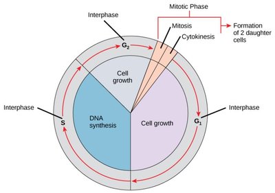
The cell cycle is the series of steps a cell undergoes to grow, replicate its DNA, and divide into two new cells. This process is fundamental for growth, tissue repair, and reproduction in all living organisms. Proper regulation of the cell cycle is essential; errors can lead to diseases such as cancer or cell death.
Phases of the Cell Cycle
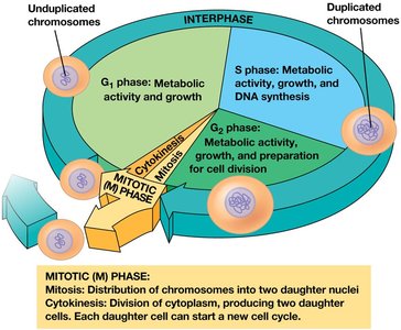
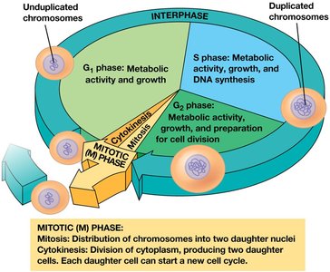
Interphase: The cell grows, performs normal functions, and prepares for division. It consists of three subphases:
G1 phase (First Gap): Cell growth and normal activity. Cells may enter a non-dividing state called G0.
S phase (Synthesis): DNA replication occurs.
G2 phase (Second Gap): Further growth and preparation for division.
Mitotic (M) phase: Includes mitosis (division of the nucleus) and cytokinesis (division of the cytoplasm), resulting in two daughter cells.
Cell Division
Definition and Types
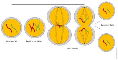
Cell division is the process by which a single cell splits to form two new cells. Before division, the cell copies its DNA so each new cell receives identical genetic information. There are two main types:
Mitosis: Produces genetically identical cells, used for growth, repair, and asexual reproduction.
Meiosis: Produces cells with half the genetic material, used for sexual reproduction.
Functions of Cell Division
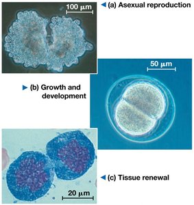
Reproduction: Single-celled organisms divide to produce new individuals.
Growth and Development: Multicellular organisms develop from a single fertilized egg through repeated cell divisions.
Tissue Repair and Renewal: Replaces damaged or worn-out cells.
Genetic Continuity: Ensures each new cell receives a complete set of DNA.
Organization of Genetic Material
Genome Structure
The genome is the complete set of genetic material in a cell. It contains all the instructions for cell function and development.
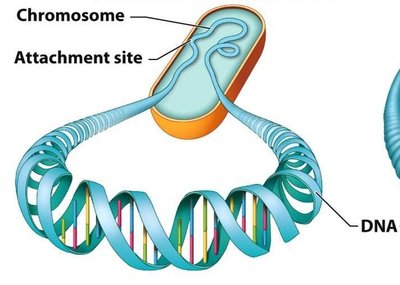
Prokaryotic cells: Usually have a single circular chromosome located in the nucleoid, and may contain plasmids.
Eukaryotic cells: Have multiple linear chromosomes located in the nucleus.
Chromatin and Chromosomes
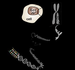
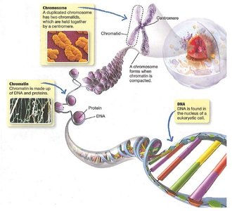
In eukaryotes, DNA is packaged with proteins to form chromatin. Chromatin condenses to form visible chromosomes during cell division. DNA wraps around histone proteins for efficient packaging.
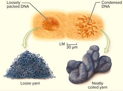
Chromatin is less condensed during interphase, allowing access for gene expression.
Chromosomes become highly condensed before division for accurate segregation.
Chromosome Sets and Ploidy
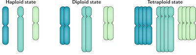
Chromosomes in eukaryotes are organized in sets:
Homologous chromosomes: Matching pairs, one from each parent.
Ploidy: Number of chromosome sets in a cell.
Haploid (n): One set
Diploid (2n): Two sets
Polyploid (>2n): More than two sets
Germ Line and Somatic Cells
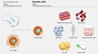
Germ line cells: Produce gametes (sperm or eggs), are haploid after meiosis.
Somatic cells: Make up most of the body, are diploid, and divide by mitosis.
Chromosome Number in Different Species
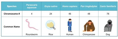
Each species has a characteristic chromosome number, usually referring to the diploid number in somatic cells. Chromosome number does not correlate with organism complexity.
Species | Chromosome # | Common Name |
|---|---|---|
Parascaris equorum | 4 | Roundworm |
Oryza sativa | 24 | Rice |
Homo sapiens | 46 | Human |
Pan troglodytes | 48 | Chimpanzee |
Canis familiaris | 78 | Dog |
Stages of Mitosis
Sequence of Events
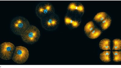
Mitosis is the process by which the nucleus and chromosomes are divided. It occurs in a precise sequence:
Prophase: Chromosomes condense, spindle forms, nucleolus disappears.
Prometaphase: Nuclear envelope breaks down, spindle fibers attach to kinetochores.
Metaphase: Chromosomes align at the metaphase plate.
Anaphase: Sister chromatids separate and move to opposite poles.
Telophase: Chromosomes decondense, nuclear envelopes reform.
Cytokinesis: Cytoplasm divides, forming two daughter cells.
Chromosome Duplication and Distribution
Before division, each chromosome is duplicated, forming two identical sister chromatids joined at the centromere. During mitosis, sister chromatids are separated so each daughter cell receives a complete set of chromosomes.
Mitotic Spindle and Centrosomes
Structure and Function
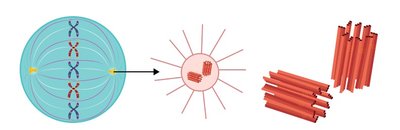
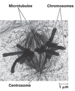
The mitotic spindle is a structure made of microtubules that organizes and separates chromosomes during mitosis. The centrosome is the main microtubule-organizing center in animal cells, containing centrioles.
Centrosomes duplicate during interphase and move to opposite poles in mitosis.
Asters help anchor the spindle.
Each sister chromatid has a kinetochore at its centromere, which attaches to spindle microtubules.
Cell Cycle Regulation
Control Mechanisms
Cell division is tightly regulated by internal and external signals. Proteins called cyclins and cyclin-dependent kinases (CDKs) control progression through the cell cycle. Checkpoints ensure the cell is ready to proceed:
G1 checkpoint: Checks DNA integrity, nutrients, and growth signals.
G2 checkpoint: Ensures DNA replication is complete and undamaged.
M checkpoint: Verifies all chromosomes are properly attached to spindle fibers before separation.
If problems are detected, the cell can pause for repair or enter a non-dividing state. If damage is irreparable, the cell may undergo apoptosis (programmed cell death).
Loss of Cell Cycle Control and Cancer
Consequences of Dysregulation
Cancer cells can bypass normal cell cycle controls, leading to uncontrolled division. They may produce their own growth signals, ignore checkpoints, evade apoptosis, and pass damaged DNA to daughter cells. Excessive division forms tumors, which can be benign (localized) or malignant (invasive). Malignant cells can spread (metastasize) to distant sites.
Prokaryotic Cell Division: Binary Fission
Process and Steps
Prokaryotes divide by binary fission, a simpler process than mitosis:
The cell copies its chromosome.
The cell elongates, and chromosome copies move apart.
The plasma membrane pinches inward, forming a new cell wall.
Two genetically identical daughter cells result.
Summary Table: Cell Cycle Phases
Phase | Main Events |
|---|---|
G1 | Cell growth, normal activity |
S | DNA synthesis (replication) |
G2 | Preparation for division |
M | Mitosis and cytokinesis |
Key Equations
Cell cycle progression is regulated by cyclin-CDK complexes:
Chromosome number in diploid cells:
Chromosome number in haploid cells:
Additional info: Academic context was added to clarify the functions of cell cycle checkpoints, the distinction between mitosis and meiosis, and the consequences of cell cycle dysregulation in cancer.
