 뒤로
뒤로The Cell Cycle, Mitosis, and Meiosis: Structure and Regulation
스터디 가이드 - 스마트 노트
 자료에 맞춘 맞춤형 노트, 핵심 정의, 예시, 맥락을 확장해 제공합니다.
The Cell Cycle
Overview of Cell Cycle Stages
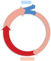
The cell cycle is a series of events that cells undergo to grow and divide. It consists of four main stages: two growth phases (G1 and G2), DNA replication (S phase), and cell division (M phase).
G1 Phase: Cells perform their normal functions and grow.
S Phase: DNA is replicated, producing two identical copies of each chromosome.
G2 Phase: Cells prepare for mitosis by growing further and replicating organelles.
M Phase (Mitosis): Chromosomes are separated and the cell divides.
Interphase and Mitosis
Cells alternate between interphase (G1, S, G2) and mitosis (M phase). Interphase is the period of growth and DNA replication, while mitosis is the process of chromosome separation and cell division. 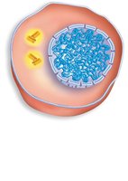
Regulation of the Cell Cycle
Cell Cycle Checkpoints
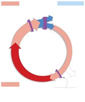
The cell cycle is tightly regulated by checkpoints that ensure proper progression and prevent errors.
G1 Checkpoint: Ensures cell size, nutrient availability, social signals, and DNA integrity. Cells that do not pass enter the G0 state.
G2 Checkpoint: Ensures successful DNA replication and undamaged DNA. Activated MPF (Maturation Promoting Factor) must be present.
M-phase Checkpoints: Ensure chromosomes are attached to the spindle apparatus and properly segregated. MPF must be absent for progression.
Chromosome Structure and DNA Replication
Changes to DNA During S Phase
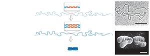
During the S phase, chromosomes are replicated, resulting in two sister chromatids joined at the centromere. Chromosomes condense before mitosis, making them visible under a microscope.
Unreplicated Chromosome: Contains a single DNA molecule.
Replicated Chromosome: Contains two identical sister chromatids.
Condensed Chromosome: Chromatids are tightly packed for mitosis.
Why Cells Divide
Functions of Cell Division
Cell division is essential for reproduction, growth, and repair.
Reproduction: Produces gametes (sexual) or new cells (asexual).
Growth: Allows organisms to develop from a single cell to multicellular forms.
Repair/Renewal: Replaces cells lost to wear, tear, or injury.
Mitosis: Steps and Mechanisms
Phases of Mitosis
Mitosis is divided into several phases, each with distinct events:
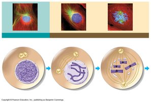
Prophase: Chromosomes condense, spindle apparatus forms, centrioles move to poles, nuclear envelope begins to fragment.
Prometaphase: Nuclear envelope fragments, microtubules attach to kinetochores, chromosomes begin moving.
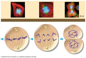
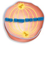
Metaphase: Chromosomes align at the metaphase plate, spindle fibers coordinate movement.
Anaphase: Sister chromatids separate and move to opposite poles, pulled by motor proteins.
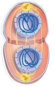
Telophase: Chromosomes arrive at poles, nuclear envelope reforms, chromosomes de-condense, spindle disperses.
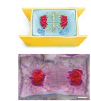
Cytokinesis: Division of cytoplasm; actin-myosin ring pinches animal cells, cell plate forms in plant cells.
Microtubules and Chromosome Movement
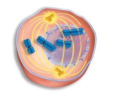
Microtubules, originating from centrosomes, coordinate chromosome movement during mitosis. They attach to kinetochores and exert forces to separate chromatids. 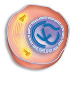
Cytokinesis in Animals and Plants
Animal Cells: Actin-myosin ring constricts, pinching the cell into two.
Plant Cells: Vesicles from Golgi fuse at the center to form a cell plate, which grows to divide the cell.
Cell Division in Bacteria
Binary Fission
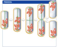
Bacteria divide by binary fission, a simpler process than mitosis.
DNA is copied and attached to the cell membrane.
Copies separate as the cell elongates.
A protein ring constricts the cell membrane.
The cell splits into two identical daughter cells.
Meiosis and Sexual Reproduction
Meiosis: Reductive Cell Division
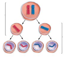
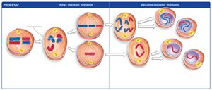
Meiosis is a two-step division process that reduces chromosome number by half, producing haploid gametes.
Meiosis I: Homologous chromosomes separate, resulting in two haploid cells.
Meiosis II: Sister chromatids separate, resulting in four genetically distinct haploid cells.
Genetic Diversity in Sexual Reproduction
Crossing-over and independent assortment during meiosis increase genetic diversity.
Fertilization combines chromosomes from two parents, further increasing variation.
Asexual reproduction produces genetically identical offspring.
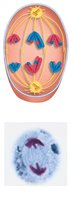
Meiosis I: Key Events
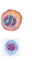
Early Prophase I: Chromosomes condense, spindle forms, synapsis occurs, forming bivalents.
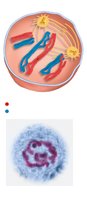
Late Prophase I: Nuclear envelope fragments, spindle attaches to kinetochores, chiasmata form, crossing-over occurs.
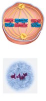
Metaphase I: Bivalents align at metaphase plate, independent assortment occurs.
Anaphase I: Homologs separate, chiasmata break.
Telophase I/Cytokinesis: Chromosomes reach poles, cell divides into two haploid cells.
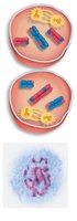
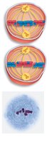
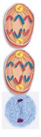
Meiosis II: Key Events
Prophase II: Spindle reforms, microtubules attach to kinetochores.
Metaphase II: Chromosomes align at metaphase plate.
Anaphase II: Sister chromatids separate.
Telophase II/Cytokinesis: Chromatids reach poles, nuclear envelope reforms, cell divides.
Errors in Meiosis
Nondisjunction and Chromosomal Abnormalities
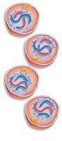
Errors such as nondisjunction can occur when homologs fail to separate, resulting in abnormal chromosome numbers in daughter cells.
Half of the daughter cells may have two copies of a chromosome, while the other half may lack it.
Such errors are common and often lead to spontaneous termination of conceptions.
Only a few chromosomal abnormalities are viable.
Summary Table: Comparison of Mitosis and Meiosis
Feature | Mitosis | Meiosis |
|---|---|---|
Number of Divisions | 1 | 2 |
Chromosome Number in Daughter Cells | Same as parent (diploid) | Half of parent (haploid) |
Genetic Identity | Identical to parent | Genetically distinct |
Function | Growth, repair, asexual reproduction | Sexual reproduction |
Additional info: Academic context was added to clarify the phases, checkpoints, and mechanisms of cell division, as well as the importance of genetic diversity and errors in meiosis.
