 뒤로
The Cell Cycle: Structure, Function, and Regulation
스터디 가이드 - 스마트 노트
 자료에 맞춘 맞춤형 노트, 핵심 정의, 예시, 맥락을 확장해 제공합니다.
The Cell Cycle
Key Roles of Cell Division
Cell division is fundamental for life, enabling organisms to reproduce, grow, and repair tissues. In unicellular organisms, division of one cell reproduces the entire organism. In multicellular eukaryotes, cell division is essential for development from a fertilized egg, growth, and tissue repair.
Asexual reproduction: Single-celled organisms reproduce by cell division.
Growth and development: Multicellular organisms grow by increasing cell number.
Tissue renewal: Damaged or old cells are replaced by new cells.
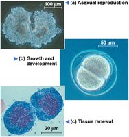
Chromosomes and Genetic Information
DNA molecules in a cell are packaged into chromosomes. Eukaryotic chromosomes consist of chromatin, a complex of DNA and protein that condenses during cell division. Most cell division results in genetically identical daughter cells.
Somatic cells: Nonreproductive cells with two sets of chromosomes.
Gametes: Reproductive cells (sperm and eggs) with half as many chromosomes as somatic cells.
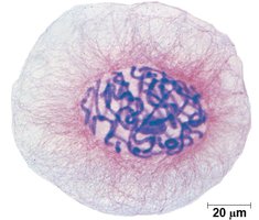
Chromosome Duplication and Distribution
Before cell division, DNA is replicated and chromosomes condense. Each duplicated chromosome has two sister chromatids attached by cohesins. The centromere is the region where chromatids are most closely attached.
Sister chromatids: Identical copies of a chromosome joined together.
Centromere: The constricted region joining sister chromatids.
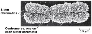
Chromosome Duplication and Distribution During Cell Division
Chromosome duplication and separation ensure each daughter cell receives a complete set of genetic information.
Chromosome duplication produces two sister chromatids.
Separation of sister chromatids during cell division distributes genetic material equally.
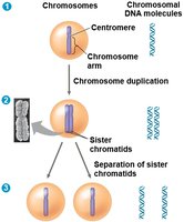
Phases of the Cell Cycle
Overview of Cell Cycle Phases
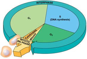
The cell cycle consists of two main phases: the Mitotic (M) phase and Interphase. The M phase includes mitosis (division of genetic material) and cytokinesis (division of cytoplasm). Interphase is subdivided into G1, S, and G2 phases.
G1 phase: Cell growth.
S phase: DNA synthesis and chromosome duplication.
G2 phase: Preparation for mitosis.
M phase: Mitosis and cytokinesis.
Mitosis: Stages and Process
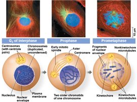
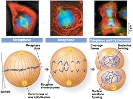
Mitosis is conventionally broken down into five stages: prophase, prometaphase, metaphase, anaphase, and telophase. Each stage is characterized by specific events that ensure accurate chromosome segregation.
Prophase: Chromosomes condense, spindle forms.
Prometaphase: Nuclear envelope fragments, spindle microtubules attach to kinetochores.
Metaphase: Chromosomes align at the metaphase plate.
Anaphase: Sister chromatids separate and move to opposite poles.
Telophase: Nuclear envelope reforms, chromosomes decondense.
The Mitotic Spindle
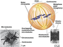
The mitotic spindle is a structure made of microtubules that controls chromosome movement during mitosis. In animal cells, spindle assembly begins in the centrosome. The spindle includes centrosomes, spindle microtubules, and asters.
Kinetochores: Protein complexes at centromeres where spindle microtubules attach.
Metaphase plate: Imaginary plane where chromosomes align during metaphase.
Cytokinesis
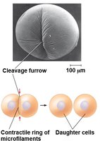
Cytokinesis is the division of the cytoplasm, resulting in two daughter cells. In animal cells, cytokinesis occurs by cleavage, forming a cleavage furrow.
Cleavage furrow: Indentation that begins the process of splitting the cell.
Contractile ring: Microfilaments that pinch the cell in two.
Regulation of the Cell Cycle
The Cell Cycle Control System
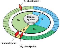
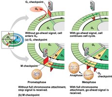
The eukaryotic cell cycle is regulated by a molecular control system. This system includes checkpoints at G1, G2, and M phases, where the cell cycle can stop until a go-ahead signal is received.
Checkpoints: Control points where the cell cycle is regulated.
G1 checkpoint: Determines if the cell will proceed to DNA synthesis.
G2 checkpoint: Ensures DNA is properly replicated before mitosis.
M checkpoint: Ensures chromosomes are properly attached to the spindle before separation.
Stop and Go Signs: Internal and External Signals
Cells respond to internal and external signals at checkpoints. If a cell receives a go-ahead signal, it completes the cell cycle and divides. If not, it exits the cycle and enters a nondividing state (G0 phase).
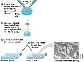
Growth factors: Stimulate cell division (e.g., PDGF).
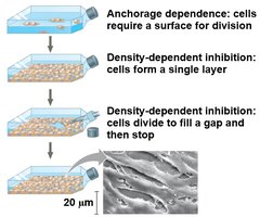
Anchorage dependence: Cells must be attached to a substratum to divide.
Density-dependent inhibition: Crowded cells stop dividing.
Growth Factors and Cell Division
Growth factors, such as Platelet-derived Growth Factor (PDGF), are released by certain cells and stimulate other cells to divide. PDGF is made by platelets and is essential for wound healing.
Density-dependent Inhibition and Anchorage Dependence
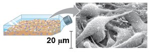
Normal mammalian cells exhibit anchorage dependence and density-dependent inhibition. Cancer cells lose these controls, leading to uncontrolled growth.
Loss of Cell Cycle Controls in Cancer Cells
Cancer and Cell Cycle Regulation
Cancer cells do not respond normally to the body’s control mechanisms. They may produce their own growth factors, convey growth factor signals without the presence of the factor, or have abnormal cell cycle control systems.
Transformation: Cells acquire the ability to divide indefinitely.
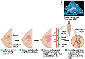
Tumors: Masses of abnormal cells within normal tissue.
Benign tumor: Abnormal cells remain at the original site.
Malignant tumor: Invades surrounding tissues and can undergo metastasis.
Metastasis: Spread of cancer cells to other parts of the body.
Cancer Treatment and Research
Metastatic tumors are treated with chemotherapeutic drugs that target the cell cycle. Side effects are due to effects on normal cells that divide frequently. Advances in molecular techniques are leading to more personalized cancer treatments.
Summary Table: Cell Cycle Phases and Checkpoints
Phase | Main Event | Checkpoint |
|---|---|---|
G1 | Cell growth | G1 checkpoint: DNA integrity, growth signals |
S | DNA synthesis | None |
G2 | Preparation for mitosis | G2 checkpoint: DNA replication completion |
M | Mitosis and cytokinesis | M checkpoint: Chromosome attachment to spindle |
Additional info: The cell cycle is tightly regulated to ensure proper growth, development, and maintenance of tissues. Disruption of these controls can lead to diseases such as cancer.
