 뒤로
The Cell Cycle: Structure, Regulation, and Cancer
스터디 가이드 - 스마트 노트
 자료에 맞춘 맞춤형 노트, 핵심 정의, 예시, 맥락을 확장해 제공합니다.
The Cell Cycle
Overview and Phases
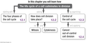
The cell cycle is the series of events that cells undergo to grow and divide. It is essential for development, tissue maintenance, and reproduction in eukaryotes. The cycle consists of four main phases: G1, S, G2, and M phase.
G1 phase: Cell grows and prepares for DNA replication.
S phase: DNA synthesis occurs, resulting in chromosome replication.
G2 phase: Cell prepares for mitosis.
M phase: Includes mitosis (nuclear division) and cytokinesis (cytoplasmic division).
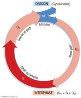
Additional info: Interphase (G1, S, G2) is when the cell is not actively dividing but is metabolically active and preparing for division.
Chromosome Structure and Replication
Chromosomes and Chromatids
Chromosomes are long, double helices of DNA wrapped around histone proteins. Each chromosome contains genes, which are regions of DNA coding for specific RNAs and proteins.
Unreplicated chromosome: Single DNA double helix.
Replicated chromosome: Two identical DNA double helices (sister chromatids) joined at the centromere.
Condensed replicated chromosome: Chromatids are tightly packed for mitosis.
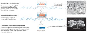
Sister chromatids are the result of DNA replication and remain attached at the centromere until separated during mitosis.
Cell Cycle Progression
Interphase and M Phase
Cells alternate between interphase (growth and preparation) and M phase (division). Most cells spend the majority of their time in interphase.
Interphase: Chromosomes are uncoiled; cell grows and prepares for division.
M phase: Chromosomes condense and are separated into daughter cells.
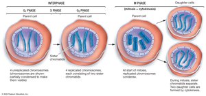
Mitosis: Process and Subphases
Stages of Mitosis
Mitosis is a continuous process divided into five subphases:
Prophase: Chromosomes condense; spindle apparatus forms.
Prometaphase: Nuclear envelope breaks down; microtubules attach to kinetochores.
Metaphase: Chromosomes align at the metaphase plate.
Anaphase: Sister chromatids are pulled apart to opposite poles.
Telophase: Nuclear envelope reforms; chromosomes decondense.
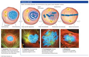
Additional info: The spindle apparatus is made of microtubules and is essential for chromosome movement.
Chromosome Movement during Anaphase
Kinetochore Microtubules
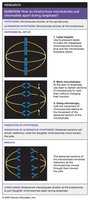
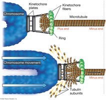
During anaphase, kinetochore microtubules shorten, pulling chromatids toward opposite poles. Motor proteins and microtubule disassembly drive this movement.
Kinetochore: Protein complex at the centromere where microtubules attach.
Microtubule disassembly: Tubulin subunits are lost from the plus end, moving the chromosome.
Cytokinesis
Division of Cytoplasm
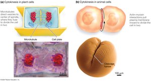
Cytokinesis is the process by which the cytoplasm divides, forming two daughter cells. The mechanism differs between plant and animal cells.
Plant cells: Vesicles form a cell plate that becomes the new cell wall.
Animal cells: Actin and myosin filaments contract to form a cleavage furrow.
Bacterial Cell Division
Binary Fission
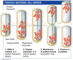
Bacteria replicate by binary fission, a process similar to eukaryotic cell division but simpler.
DNA is copied.
Copies are separated by protein filaments.
Membrane is pulled inward by a protein ring.
Fission completes the division.
Regulation of the Cell Cycle
Cell-Cycle Regulatory Molecules
The cell cycle is tightly regulated by proteins such as cyclins and cyclin-dependent kinases (Cdks). The M-phase-promoting factor (MPF) is a key regulator.
Cyclin: Regulatory protein whose concentration fluctuates during the cell cycle.
Cdk: Protein kinase active only when bound to cyclin.
MPF: Cyclin-Cdk complex that triggers entry into M phase.
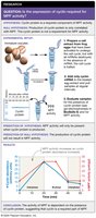
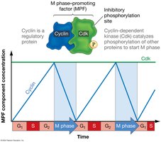
Additional info: MPF activity is regulated by phosphorylation and dephosphorylation events.
Cell-Cycle Checkpoints
G1, G2, and M-Phase Checkpoints
Checkpoints ensure that cells only proceed through the cycle when conditions are appropriate.
G1 checkpoint: Checks cell size, nutrients, DNA damage, and social signals.
G2 checkpoint: Ensures DNA replication is complete and undamaged.
M-phase checkpoints: Ensure proper chromosome attachment and separation.
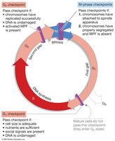
p53 protein is a tumor suppressor that can halt the cell cycle or trigger apoptosis if DNA is damaged.
Cancer and Cell Cycle Control
Loss of Regulation and Tumor Formation
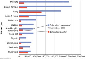
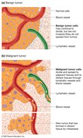
Cancer results from uncontrolled cell division due to defects in cell-cycle regulation. Tumors can be benign (noninvasive) or malignant (invasive and metastatic).
Defects in growth regulation: Activation of growth proteins or loss of tumor suppressors.
Malignant tumors: Can spread via blood or lymph to form secondary tumors (metastasis).
Benign tumors: Do not invade other tissues.
Additional info: The Rb protein and E2F are key regulators at the G1 checkpoint; defects in these can lead to cancer.
Summary Table: Cell Cycle Phases and Key Events
Phase | Main Event | Key Structures |
|---|---|---|
G1 | Cell growth, preparation for DNA replication | Chromatin, growth factors |
S | DNA synthesis, chromosome replication | DNA polymerase, sister chromatids |
G2 | Preparation for mitosis | Centrosomes, spindle apparatus |
M | Mitosis and cytokinesis | Spindle, kinetochores, actin/myosin |
