 뒤로
The Cytoskeleton and Eukaryotic Cell Structure: A Mini-Textbook Study Guide
스터디 가이드 - 스마트 노트
 자료에 맞춘 맞춤형 노트, 핵심 정의, 예시, 맥락을 확장해 제공합니다.
A Tour of the Cell: Eukaryotic Cell Structure and the Cytoskeleton
Overview of Eukaryotic Cell Components
Eukaryotic cells are highly organized structures containing various organelles, each with distinct functions essential for cellular life. Understanding the parts list and their roles is foundational for General Biology.
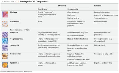
Organelle | Structure | Components | Function |
|---|---|---|---|
Nucleus | Double membrane with nuclear pores | Chromosomes, nucleolus, nuclear lamina | Genetic information storage, assembly of ribosome subunits, structural support |
Ribosomes | None | Large/small subunits, complex of RNA and proteins | Protein synthesis |
Rough ER | Single membrane with receptors | Network of branching sacs, ribosomes | Protein synthesis and processing |
Golgi apparatus | Single membrane with receptors | Stack of flattened cisternae | Protein processing (e.g., glycosylation) |
Smooth ER | Single membrane | Network of branching sacs, enzymes for lipid synthesis | Lipid synthesis |
Peroxisomes | Single membrane | Enzymes for oxidation reactions | Processing of fatty acids |
Lysosomes | Single membrane with proton pumps | Acid hydrolases | Digestion and recycling |
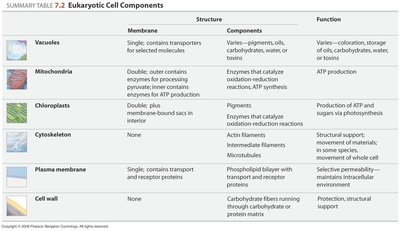
Vacuoles | Single membrane with transporters | Varies: pigments, oils, carbohydrates, water, toxins | Storage, coloration, water balance |
Mitochondria | Double membrane | Enzymes for ATP production | ATP production |
Chloroplasts | Double membrane plus internal sacs | Pigments, enzymes for photosynthesis | ATP and sugar production via photosynthesis |
Cytoskeleton | None | Actin filaments, intermediate filaments, microtubules | Structural support, movement, transport |
Plasma membrane | Single membrane with transporters | Phospholipid bilayer with proteins | Selective permeability, cell communication |
Cell wall | None | Carbohydrate fibers | Protection, structural support |
The Cytoskeleton: Structure and Function
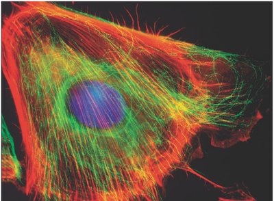
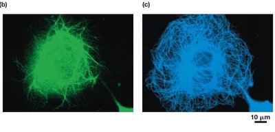
The cytoskeleton is a complex network of protein fibers that provides structural support, maintains cell shape, and enables movement and transport within the cell. It is dynamic, constantly reorganizing to meet cellular needs.
Structural Support: Maintains cell shape and anchors organelles.
Intracellular Transport: Acts as a highway for vesicle and organelle movement.
Cell Motility: Enables cell movement and division.
Major Cytoskeletal Elements
There are three primary types of cytoskeletal elements, each with unique structure and function:
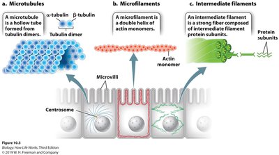
Actin Filaments (Microfilaments): Smallest, composed of actin monomers, involved in cell shape and movement.
Intermediate Filaments: Medium-sized, diverse protein composition (e.g., keratin, lamin), provide structural support.
Microtubules: Largest, hollow tubes made of tubulin dimers, involved in transport, cell division, and motility.
Actin Filaments (Microfilaments)
Actin filaments are dynamic structures that form by polymerization of actin monomers. They are typically found just beneath the plasma membrane and are crucial for maintaining cell shape and enabling movement.
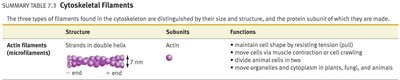
Structure: Strands in double helix, 7 nm diameter.
Functions: Resist tension, enable cell crawling, cytokinesis, and cytoplasmic streaming.
Example: Actin-myosin interactions drive muscle contraction and cell motility.
Structure | Subunits | Functions |
|---|---|---|
Strands in double helix | Actin | Maintain cell shape, muscle contraction, cell crawling, divide animal cells, move organelles/cytoplasm |
Actin-Myosin Interactions
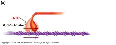
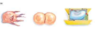
Actin filaments interact with the motor protein myosin to produce movement. This interaction is fundamental to processes such as muscle contraction, cell crawling, and cytokinesis.
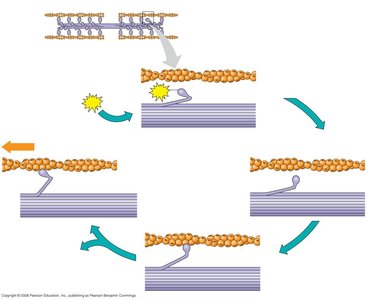
Mechanism: Myosin heads bind to actin, hydrolyze ATP, and cause actin filaments to slide.
Applications: Cell division, cytoplasmic streaming, and muscle contraction.
Intermediate Filaments
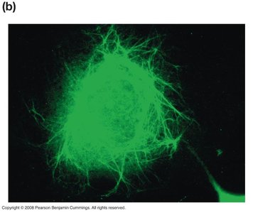
Intermediate filaments are defined by their size and diverse protein composition. They provide mechanical strength and structural support but are not involved in cell movement.
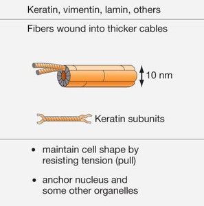
Structure: Fibers wound into thicker cables, 10 nm diameter.
Subunits: Keratin, vimentin, lamin, others.
Functions: Maintain cell shape by resisting tension, anchor nucleus and organelles.
Microtubules
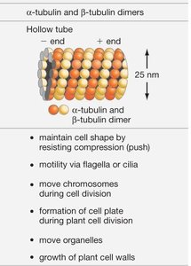
Microtubules are large, hollow tubes composed of α- and β-tubulin dimers. They are dynamic, growing and shrinking from their plus ends, and serve as tracks for intracellular transport and cell division.
Structure: Hollow tube, 25 nm diameter.
Subunits: Tubulin dimers.
Functions: Maintain cell shape by resisting compression, move chromosomes during cell division, vesicle transport, organelle arrangement, motility via cilia and flagella.
Microtubule Organizing Centers and Centrosomes
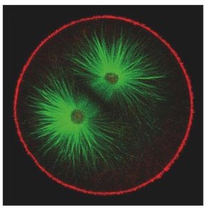
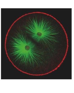
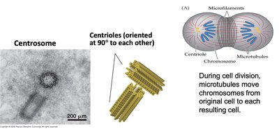
Microtubules originate from microtubule organizing centers (MTOCs), with the centrosome being the primary MTOC in animal cells. Centrosomes contain centrioles, which are essential for microtubule organization during cell division.
Motor Proteins: Kinesin and Dynein
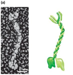
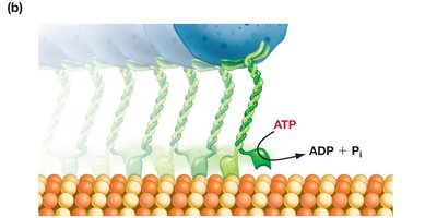
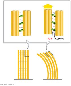
Motor proteins such as kinesin and dynein convert chemical energy from ATP into mechanical work, moving vesicles and organelles along microtubule tracks. Kinesin moves toward the plus end, while dynein moves toward the minus end.
Kinesin: Transports vesicles along microtubules, requires ATP.
Dynein: Drives movement of cilia and flagella.
Cellular Motility: Cilia and Flagella
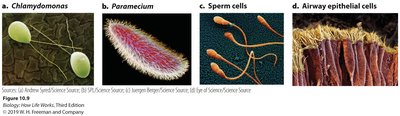
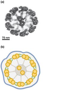
Cilia and flagella are specialized structures for cell movement, composed of microtubules arranged in a "9+2" pattern. Dynein motor proteins drive their movement by causing microtubule doublets to slide and bend.
Flagella: Long, hairlike projections for cell movement.
Cilia: Short, filament-like projections for movement or nutrient absorption.
Axoneme: Core structure with microtubules and dynein arms.
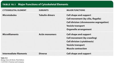
Summary Table: Major Functions of Cytoskeletal Elements
Cytoskeletal Element | Subunits | Major Functions |
|---|---|---|
Microtubules | Tubulin dimers | Cell shape/support, cell movement (cilia/flagella), cell division, vesicle transport, organelle arrangement |
Microfilaments | Actin monomers | Cell shape/support, cell movement (crawling), cell division, vesicle transport, muscle contraction |
Intermediate filaments | Diverse | Cell shape/support |
Muscle Contraction: The Sliding Filament Model
Skeletal Muscle Structure
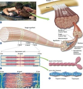
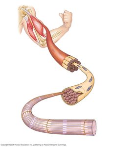
Skeletal muscle is composed of bundles of long fibers, each containing myofibrils made up of myofilaments. The functional unit of muscle contraction is the sarcomere, bordered by Z lines.
Myofilaments: Actin (thin) and myosin (thick) filaments.
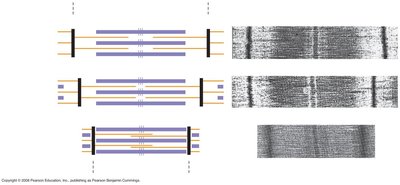
Sarcomere: Functional unit, pattern of light and dark bands.
The Sliding Filament Model
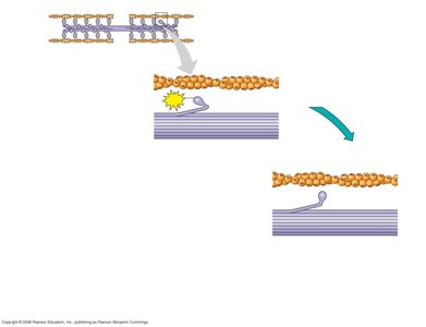
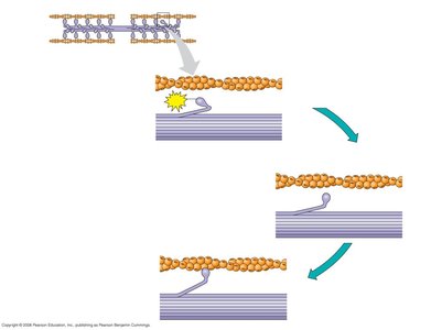
Muscle contraction occurs when actin and myosin filaments slide past each other, increasing overlap and shortening the sarcomere. Myosin heads bind to actin, hydrolyze ATP, and pull actin filaments toward the center.
ATP Hydrolysis: Provides energy for myosin head movement.
Cross-Bridge Formation: Myosin binds actin, pulls, releases, and repeats.
Key Equations and Concepts
Muscle contraction and cytoskeletal movement are powered by ATP hydrolysis:
ATP Hydrolysis:
Actin Polymerization:
Conclusion
The cytoskeleton is essential for cell structure, movement, and intracellular transport. Understanding its components and mechanisms, such as actin-myosin interactions and microtubule-based motility, is fundamental for General Biology students.
