 뒤로
뒤로Cell Junctions and Extracellular Structures: Structure, Function, and Disease
스터디 가이드 - 스마트 노트
 자료에 맞춘 맞춤형 노트, 핵심 정의, 예시, 맥락을 확장해 제공합니다.
Cell Junctions and Extracellular Structures
Overview of Cell Junctions and Tissue Organization (. outside the cell )
Cell junctions are specialized structures that connect cells to one another and to the extracellular matrix, playing critical roles in tissue formation, integrity, and communication.
These junctions are essential for multicellular organisms, enabling cells to form tissues and organs with distinct functions. - Cell-cell contact: Direct physical interactions between adjacent cells. - Cell adhesion: The process by which cells attach to each other or to the extracellular matrix. - Cell junctions: Specialized protein complexes that mediate cell-cell and cell-matrix adhesion. - Extracellular matrix (ECM): A network of proteins and polysaccharides outside cells that provides structural and biochemical support. 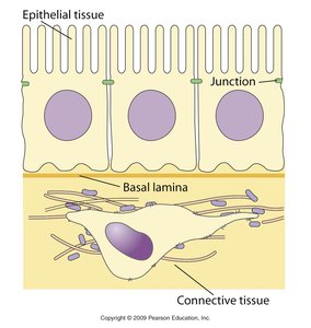
what kind of interactions ? and cell adhesion?
Types of Cell-Cell Adhesion Molecules
Cell adhesion molecules (CAMs) are proteins located on the cell surface involved in binding with other cells or with the ECM. They are classified based on their interaction types: - Homotypic (homophilic) interaction: CAMs bind to identical molecules on adjacent cells. - Heterotypic (heterophilic) interaction: CAMs bind to different molecules on adjacent cells. 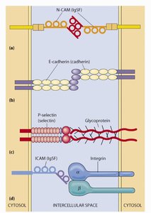
Immunoglobulin Superfamily (IgSF)
- NCAM (Neuronal Cell Adhesion Molecule): Involved in neuronal development and growth ( they grow into the tuissue- they follow chemical released by what they're looking for ) . Mutations can lead to mental retardation. 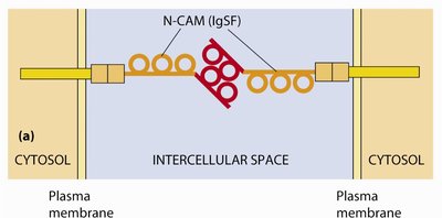
Cadherins
- E-cadherin: Expressed in epithelial cells; essential for embryonic development and tissue formation. Knockout (KO) models lack adhesion.
- N-cadherin: Found in synapses; requires Ca2+ for adhesion. 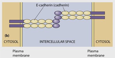
Selectins
if you eat mayo containing edta what consequences does it have ?
- When you consume commercial mayonnaise, Calcium Disodium EDTA is often included in tiny amounts as a food preservative. It acts as a chelating agent, meaning it binds to trace metal ions to prevent chemical oxidation and keep the oil and egg mixture from spoiling or separating Ethylenediaminetetraacetic acid (EDTA)
- P-selectin: Located on platelets and endothelial cells. - L-selectin: Found on leukocytes; involved in leukocyte rolling. - Glycosylation: Critical for selectin function. 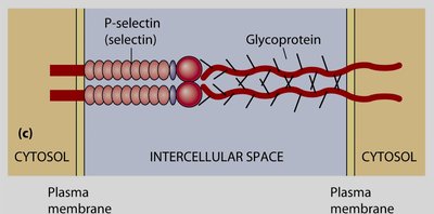
Major Types of Cell Junctions
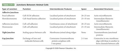
Animal cells possess several types of junctions, each with distinct functions and associated structures.
Type of Junction | Function | Intermembrane Features | Space | Associated Structures |
|---|---|---|---|---|
Focal adhesion | Cell-ECM adhesion | Localized points of attachment | 20–35 nm | Actin microfilaments |
Hemidesmosome | Cell-basal lamina adhesion | Localized points of attachment | 25–35 nm | Intermediate filaments (tonofilaments) |
Adherens junction | Cell-cell adhesion | Continuous zones of attachment | 20–35 nm | Actin microfilaments |
Desmosome | Cell-cell adhesion | Localized points of attachment | 25–35 nm | Intermediate filaments |
Tight junction | Sealing spaces between cells | Membranes joined along ridges | 0 nm | Transmembrane junctional proteins |
Gap junction | Exchange of ions and molecules between cells | Continuous transmembrane protein complexes within 3–nm pores | 2–3 nm | Connexins in one membrane align with those in another to form channels between cells |
Adhesive Junctions: Adherens Junctions and Desmosomes
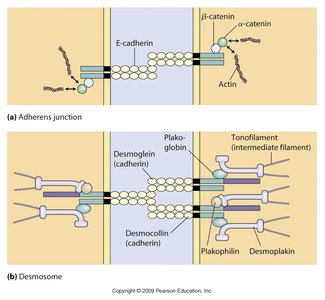
Adhesive junctions connect cells to each other and to the ECM, providing mechanical strength and signaling functions. - Adherens junctions: Link actin cytoskeletons of adjacent cells via cadherins. - Desmosomes: Provide strong adhesion through cadherins (desmoglein, desmocollin) and intermediate
filaments. 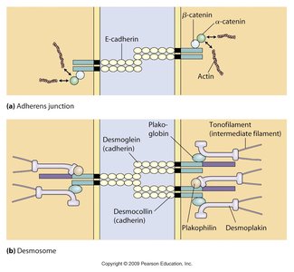
adhesion is important - in cancers
Key Components of Adherens Junctions
Cadherins: Calcium-dependent proteins on the cell surface that bind directly to identical cadherins on adjacent cells.
Catenins: Intracellular molecules (including beta-catenin, p120-catenin, and alpha-catenin) that attach the tail of the cadherin to the actin network.
Actin Filaments: The structural protein cables inside the cell that provide tension support and shape
Clinical Relevance: Naxos Disease
- Caused by mutations in plakoglobin and desmoplakin. - Characterized by woolly hair and progressive heart disease (cardiomyopathies with cutaneous * low blood pressure phenotype). 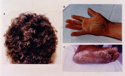
Tight Junctions
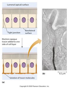
sodium go thru not CA Tight junctions seal spaces between cells, preventing the passage of molecules and ions through the space between cells. - Claudins: Key proteins in tight junctions; about 12 different types, with dynamic interactions and selectivity for cations. 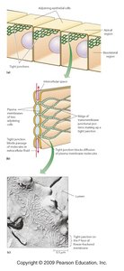
Cell Junctions in Host-Pathogen Interactions
Pathogens exploit cell adhesion proteins to invade host cells. - Yersinia: Uses integrin to invade cells, causing gastroenteritis. - Listeria: (very to treat with antibiotics Uses internalin A and E-cadherin for entry; can cause lethal bacterial meningitis. 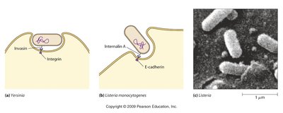
Adenoviruses interact with cell adhesion molecules and adhesion receptors in a sequential, two-step process involving distinct viral capsid proteins for initial cell attachment and secondary internalization
Extracellular Matrix (ECM) and Its Components
The ECM is a complex network of proteins and polysaccharides that provides structural support and regulates cell behavior. - Collagen: Provides strength. - Elastin: Provides flexibility. - Fibronectin: Involved in cell shape, movement, and blood clotting. - Laminins: Mediate cell attachment to connective tissue. - Integrins: Connect ECM to cytoskeleton and mediate signaling. 
Fibronectin( Fibronectin is a large, multi-functional glycoprotein found in the extracellular matrix and blood plasma that helps cells stick, move, and organize tissues
- Soluble in blood, binds fibrin, and facilitates platelet attachment. - Essential for tissue formation, cell movement, and attachment. 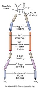
Laminin(a vital family of structural glycoproteins found in the basement membranes of virtually all animal tissues )
-
Present in basal lamina; defects cause muscular dystrophy, intestinal, and kidney disease. 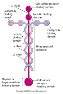
Integrins
- Mediate focal adhesion and inside-out signaling. - Required for anchorage-dependent growth. 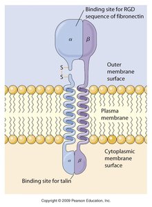
Integrins ( inside out signal - is it gonna work or release it- cell regulates its attachment (transmembrane receptor proteins that enable cells to attach to the extracellular matrix and communicate with their surrounding environment)
Collagen
- With age, collagen fibers- fibrils become crosslinked and inflexible. - Defects in Collagen I and III cause Ehlers-Danlos Syndrome.
Elastin
- Essential for wound healing and skin elasticity. - Loss of elastin with age leads to skin changes.
-defect: Marfan Syndrome: Characterized by hypermobility and unique physical features. Example: Paganini's hypermobility and Harry Houdini's flexibility are attributed to connective tissue properties.
Summary Table: Major ECM Proteins and Their Functions
Protein | Function | Clinical Relevance |
|---|---|---|
Collagen | Strength | Ehlers-Danlos Syndrome |
Elastin | Flexibility (on the joints) w age: loss elastin - wound heal. | Marfan Syndrome |
Fibronectin | Cell shape, movement, blood clotting | Wound healing |
Laminin | Cell attachment to connective tissue | Muscular dystrophy, kidney disease |
Integrin | Connect ECM and cytoskeleton | Cell signaling, cancer |
Additional info: Expanded explanations and clinical context were added to ensure completeness and academic quality.
