 뒤로
Cytoskeletal Systems and Cellular Motility: Microtubules, Microfilaments, and Intermediate Filaments
스터디 가이드 - 스마트 노트
 자료에 맞춘 맞춤형 노트, 핵심 정의, 예시, 맥락을 확장해 제공합니다.
Cytoskeleton and Cellular Motility
Overview of Cytoskeletal Functions
The cytoskeleton is a dynamic network of protein filaments that provides structural support, facilitates intracellular transport, and enables cell movement. It is essential for maintaining cell shape, polarity, and the organization of organelles.
Chromosome movements: The cytoskeleton is crucial during mitosis and meiosis for the proper segregation of chromosomes.
Maintaining shape and polarity: Cytoskeletal elements determine cell morphology and spatial organization.
Trafficking organelles: Motor proteins move vesicles and organelles along cytoskeletal tracks.
Cell motility: Cells move by reorganizing their cytoskeleton, important in processes like wound healing and immune response.
Muscle contraction: Actin and myosin filaments interact to produce contraction.
Cell locomotion: Cells crawl or swim using cytoskeletal structures.
Cytoplasmic streaming: Movement of cytoplasm facilitated by cytoskeletal elements.
Nuclear scaffolding: Intermediate filaments provide structural support to the nucleus.
Neurofilaments: Specialized intermediate filaments in neurons maintain axonal structure.
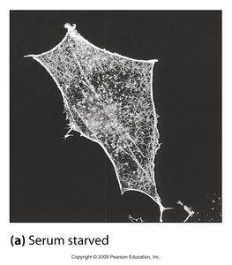
Properties of Cytoskeletal Filaments
Microtubules, Microfilaments, and Intermediate Filaments
Cytoskeletal filaments are classified into three main types, each with distinct properties and functions.
Microtubules: Hollow tubes composed of tubulin dimers, involved in cell division, intracellular transport, and structural support.
Microfilaments: Thin filaments made of actin, important for cell shape, movement, and muscle contraction.
Intermediate filaments: Rope-like structures providing mechanical strength and nuclear scaffolding.
Filament | Polymer Structure | Subunit | Diameter | Function |
|---|---|---|---|---|
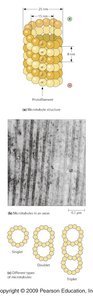
Microtubules | Hollow tube of 13 protofilaments | α/β-tubulin dimer | 25 nm | Cell shape, transport, mitosis |
Microfilaments | Two intertwined chains | Actin monomer | 7 nm | Cell shape, motility, contraction |
Intermediate Filaments | Eight protofilaments | Varied (e.g., keratin, vimentin) | 8–12 nm | Mechanical strength, nuclear support |

Microtubule Structure and Assembly
Microtubule Organization
Microtubules are cylindrical polymers of tubulin, organized as singlets, doublets, or triplets depending on their cellular location.
Singlet: Most cytoplasmic microtubules.
Doublet: Found in cilia and flagella.
Triplet: Present in basal bodies and centrioles.
Microtubule Assembly
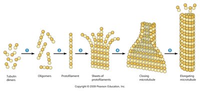
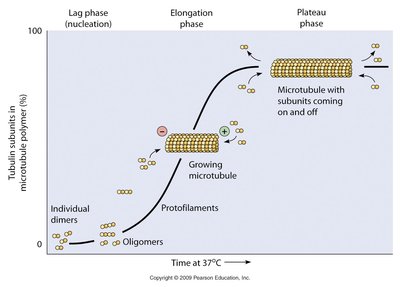
Microtubule polymerization is a multi-step process involving nucleation, elongation, and plateau phases.
Nucleation: Tubulin dimers form oligomers, which assemble into protofilaments.
Elongation: Sheets of protofilaments close to form a cylindrical microtubule.
Plateau: Microtubule reaches steady-state with subunits coming on and off.
Microtubule Polarity and Treadmilling
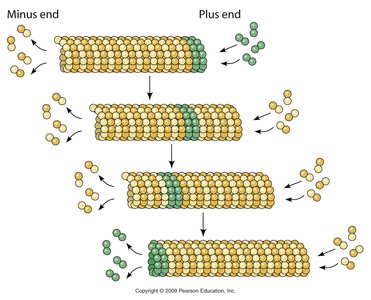
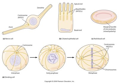
Microtubules are polar structures with distinct plus and minus ends, influencing their dynamics and cellular functions.
Plus end: Rapid growth and shrinkage.
Minus end: Usually anchored at microtubule organizing centers (MTOCs).
Treadmilling: Subunits are added at the plus end and removed at the minus end, resulting in dynamic instability.
Drugs Affecting the Cytoskeleton
Pharmacological Perturbation
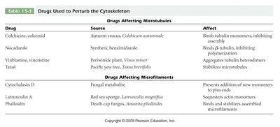
Several drugs can disrupt cytoskeletal function by targeting microtubules or microfilaments.
Colchicine, nocodazole: Inhibit microtubule assembly.
Vinblastine, vincristine: Inhibit microtubule polymerization.
Taxol: Stabilizes microtubules.
Cytochalasin D, latrunculin A, phalloidin: Affect actin microfilaments by preventing polymerization or stabilizing filaments.
Drug | Source | Affect |
|---|---|---|
Colchicine, colcemid | Autumn crocus | Binds tubulin monomers, inhibiting assembly |
Nocodazole | Synthetic | Binds β-tubulin, inhibiting polymerization |
Vinblastine, vincristine | Periwinkle plant | Aggregates tubulin heterodimers |
Taxol | Pacific yew tree | Stabilizes microtubules |
Cytochalasin D | Fungal metabolite | Prevents addition of new monomers to plus end |
Latrunculin A | Red sea sponge | Sequesters actin monomers |
Phalloidin | Death cap fungus | Binds and stabilizes assembled microfilaments |
Microfilaments (Actin Filaments)
Structure and Function
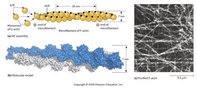
Microfilaments are composed of actin and are essential for cell shape, motility, and muscle contraction.
Actin monomers: Polymerize to form F-actin filaments.
ATP hydrolysis: Drives filament assembly and dynamics.
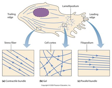
Cell cortex: Dense network of actin beneath the plasma membrane.
Actin Filaments in Crawling Cells
Crawling cells utilize actin filaments to form structures such as lamellipodia and filopodia, enabling movement.
Lamellipodium: Broad, sheet-like protrusion at the leading edge.
Filopodium: Thin, spike-like projections.
Stress fibers: Contractile bundles of actin.

Actin-Binding Proteins
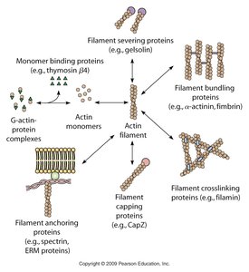
Actin-binding proteins regulate the organization, dynamics, and function of actin filaments.
Filament severing proteins: Gelsolin severs actin filaments.
Filament bundling proteins: α-actinin, fimbrin bundle actin filaments.
Filament crosslinking proteins: Filamin crosslinks actin filaments.
Filament capping proteins: CapZ caps the ends of actin filaments.
Filament anchoring proteins: Spectrin, ERM proteins anchor actin to membranes.
Intermediate Filaments
Structure and Assembly
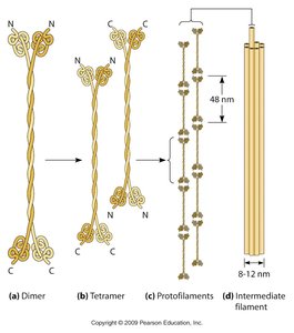
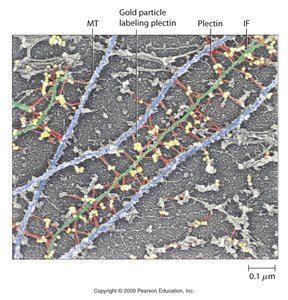
Intermediate filaments are composed of diverse proteins and provide mechanical strength to cells and tissues.
Dimer: Two polypeptides form a coiled-coil.
Tetramer: Two dimers associate in an antiparallel fashion.
Protofilament: Tetramers assemble into protofilaments.
Intermediate filament: Eight protofilaments form a mature filament.
Classes and Functions of Intermediate Filaments
Intermediate filaments are classified based on their protein composition and tissue distribution.
Class I & II: Keratins in epithelial cells for mechanical strength.
Class III: Vimentin, desmin, and others in mesenchymal and muscle cells.
Class IV: Neurofilaments in neurons for axon structure.
Class V: Nuclear lamins in all cell types for nuclear scaffolding.
Class VI: Nestin in neural stem cells.
Class | IF Protein | Molecular Mass (kDa) | Tissue | Function |
|---|---|---|---|---|
I | Acidic cytokeratins | 40–56 | Epithelial cells | Mechanical strength |
II | Basic cytokeratins | 57–67 | Epithelial cells | Mechanical strength |
III | Vimentin, desmin | 53–54 | Mesenchymal, muscle cells | Cell shape, contractile machinery |
IV | Neurofilaments | 62–110 | Neurons | Axon structure |
V | Lamin A, B, C | 70 | All cell types | Nuclear scaffolding |
VI | Nestin | 240 | Neural stem cells | Unknown |
Summary
The cytoskeleton is a complex and dynamic system essential for cell structure, function, and movement. Microtubules, microfilaments, and intermediate filaments each play unique roles in cellular processes, and their regulation is critical for normal cell physiology and response to environmental changes. Understanding the cytoskeleton is fundamental for cell biology, as it underlies many aspects of cell behavior, development, and disease.
