 뒤로
뒤로Mitosis and Meiosis: Chromosome Structure, Cell Cycle, and Genetic Variation
스터디 가이드 - 스마트 노트
 자료에 맞춘 맞춤형 노트, 핵심 정의, 예시, 맥락을 확장해 제공합니다.
Mitosis and Meiosis
Introduction to Cell Division
Mitosis and meiosis are two fundamental processes of cell division in eukaryotic organisms. Mitosis results in the production of genetically identical cells, while meiosis leads to the formation of gametes with half the chromosome number, introducing genetic diversity.
Chromosomes and Chromosome Structure
Chromosomes, Chromatin, and Chromatids
Chromosomes are highly organized structures composed of DNA and proteins, visible during cell division. Chromatin refers to the less condensed form of genetic material found during interphase. Each duplicated chromosome consists of two sister chromatids joined at a centromere.
Chromosome: A single, long DNA molecule with associated proteins.
Chromatin: The complex of DNA and proteins in the nucleus when the cell is not dividing.
Sister Chromatids: Identical copies of a chromosome connected by a centromere after DNA replication.
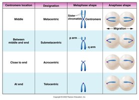
Centromere Position and Chromosome Types
Chromosomes are classified based on the position of their centromere:
Centromere Location | Designation | Metaphase Shape | Anaphase Shape |
|---|---|---|---|
Middle | Metacentric | V-shaped | Even migration |
Between middle and end | Submetacentric | L-shaped | Uneven migration |
Close to end | Acrocentric | J-shaped | Distinct migration |
At end | Telocentric | Rod-shaped | Terminal migration |
Chromosome Number: Haploid and Diploid
Organisms can be haploid (n) or diploid (2n), referring to the number of chromosome sets. Diploid cells contain homologous pairs, while haploid cells have one set. The diploid number (2n) is characteristic for each species.
Homologous Chromosomes: Chromosome pairs with the same genes but possibly different alleles.
Locus (plural: loci): The specific location of a gene on a chromosome.
Allele: Different forms of a gene found at the same locus.
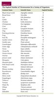
Common Name | Scientific Name | Diploid Number |
|---|---|---|
Fruit fly | Drosophila melanogaster | 8 |
Human | Homo sapiens | 46 |
Dog | Canis familiaris | 78 |
Mitosis and the Cell Cycle
Phases of the Cell Cycle
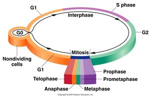
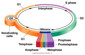
The cell cycle consists of interphase (G1, S, G2) and the mitotic phase (M). Interphase is the period of cell growth and DNA replication, while mitosis is the process of nuclear division.
G1 phase: Cell growth and preparation for DNA synthesis.
S phase: DNA replication occurs.
G2 phase: Preparation for mitosis.
M phase: Mitosis and cytokinesis.
Duration of Mitosis and Its Stages
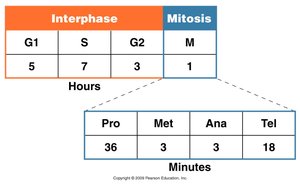
The cell cycle is tightly regulated, and the duration of each phase can vary. In a typical mammalian cell:
Phase | Duration |
|---|---|
G1 | 5 hours |
S | 7 hours |
G2 | 3 hours |
Mitosis (M) | 1 hour |
Mitosis Stage | Duration (minutes) |
|---|---|
Prophase | 36 |
Metaphase | 3 |
Anaphase | 3 |
Telophase | 18 |
Stages of Mitosis
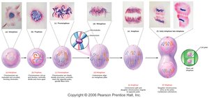
Mitosis is divided into several stages, each with distinct events:
Prophase: Chromosomes condense, spindle forms, nuclear envelope breaks down.
Metaphase: Chromosomes align at the metaphase plate.
Anaphase: Sister chromatids separate and move to opposite poles.
Telophase: Nuclear envelopes reform, chromosomes decondense.
Cytokinesis: Division of the cytoplasm, resulting in two daughter cells.
Regulation of the Cell Cycle
Cell cycle progression is controlled by cyclins and cyclin-dependent kinases (Cdks). Phosphorylation of target proteins by these complexes ensures proper timing and fidelity of cell division.
Cyclins: Regulatory proteins whose levels fluctuate during the cell cycle.
Cdk: Enzymes that, when bound to cyclins, phosphorylate other proteins to drive cell cycle transitions.
Phosphorylation: Addition of a phosphate group, often regulating protein activity.
Meiosis and Sexual Reproduction
Overview of Meiosis
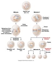
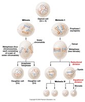
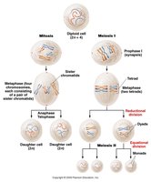
Meiosis is a specialized form of cell division that reduces the chromosome number by half, producing four genetically unique haploid cells (gametes). It consists of two sequential divisions: meiosis I (reductional) and meiosis II (equational).
Reductional Division: Homologous chromosomes separate, reducing chromosome number by half.
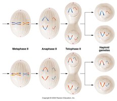
Equational Division: Sister chromatids separate, similar to mitosis.
Genetic Variation in Meiosis
Meiosis introduces genetic variation through independent assortment of chromosomes and crossing over during prophase I. Fertilization further increases genetic diversity.
Independent Assortment: Random distribution of maternal and paternal chromosomes to gametes.
Crossing Over: Exchange of genetic material between homologous chromosomes.
Stages of Meiosis I
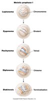
Meiosis I includes four main stages, with prophase I subdivided into five substages:
Prophase I: Homologous chromosomes pair and exchange segments (crossing over).
Metaphase I: Tetrads align at the metaphase plate.
Anaphase I: Homologous chromosomes separate (reductional division).
Telophase I: Two haploid cells form, each with duplicated chromosomes.
Prophase I Substages
Leptonema: Chromosomes condense, homology search begins.
Zygonema: Synapsis starts, synaptonemal complex forms.
Pachynema: Synapsis completes, crossing over occurs.
Diplonema: Homologs begin to separate, chiasmata visible.
Diakinesis: Terminalization of chiasmata, chromosomes prepare for metaphase I.
Synaptonemal Complex and Recombination
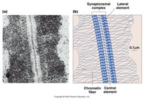
The synaptonemal complex is a protein structure essential for synapsis and crossing over during prophase I. Recombination nodules are sites of genetic exchange.
Hot Spots: Regions with high recombination frequency.
Interference: The phenomenon where one crossover event reduces the likelihood of another nearby.
Meiosis II: Equational Division
Meiosis II resembles mitosis, where sister chromatids separate, resulting in four haploid cells. This division does not reduce chromosome number further.
Comparison: Mitosis vs. Meiosis
Key Differences
Mitosis: Produces two genetically identical diploid cells; used for growth and repair.
Meiosis: Produces four genetically unique haploid cells; used for sexual reproduction.
Reduction: Chromosome number is halved in meiosis but not in mitosis.
Genetic Variation: Meiosis introduces variation through crossing over and independent assortment.
Variation in Meiosis
Sources of Genetic Diversity
Independent Assortment: Each gamete receives a random mix of maternal and paternal chromosomes.
Crossing Over: New allele combinations are created during prophase I.
Fertilization: Combines genetic material from two parents, further increasing diversity.
Gametogenesis: Spermatogenesis vs. Oogenesis
Formation of Gametes
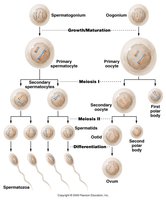
Spermatogenesis and oogenesis are the processes by which sperm and eggs are produced, respectively. Both involve meiosis but differ in timing, outcome, and cytoplasmic division.
Spermatogenesis: Produces four functional sperm cells from each primary spermatocyte.
Oogenesis: Produces one functional ovum and polar bodies from each primary oocyte.
Review for Mendelian Genetics
Transition to Mendelian Principles
The understanding of mitosis and meiosis provides the foundation for Mendelian genetics, including the behavior of alleles during gamete formation and fertilization. Key concepts include the segregation and independent assortment of alleles, which are explained by the mechanics of meiosis.
Punnett Squares: Visual tools for predicting genetic crosses.
Forked Line (Branched Diagram): Used for complex crosses involving multiple genes.
Probability Rules: Product law, sum rule, conditional probabilities, and binomial expansion are used to calculate expected outcomes of genetic crosses.
Additional info: For further study, see Mendelian genetics and probability calculations in genetic analysis.
