 뒤로
UM Exam 2 Lecture 11: Enzyme Regulation, Inhibition, and Vitamins: Key Concepts for GOB Chemistry
스터디 가이드 - 스마트 노트
 자료에 맞춘 맞춤형 노트, 핵심 정의, 예시, 맥락을 확장해 제공합니다.
Enzyme Inhibition
Types of Enzyme Inhibition
Enzyme inhibition is a critical concept in biochemistry, describing how molecules can decrease or halt enzyme activity. There are several types of inhibition, each affecting enzyme function differently.
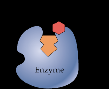
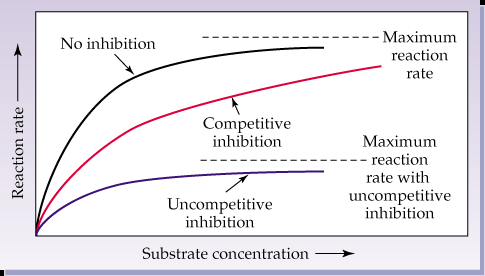
Competitive Inhibition: The inhibitor resembles the substrate and competes for the same binding site on the enzyme. This type of inhibition increases the apparent Km (substrate concentration at half-maximal velocity) but does not affect Vmax (maximum reaction rate).
Noncompetitive Inhibition: The inhibitor binds to a site other than the active site, causing a change in enzyme configuration. This decreases Vmax but leaves Km unchanged.
Uncompetitive Inhibition: The inhibitor binds only to the enzyme-substrate complex, not to the free enzyme. Both Km and Vmax are lowered by the same ratio.
Irreversible Inhibition: The inhibitor covalently modifies the enzyme, rendering it permanently inactive.
Example: Sarin, a nerve agent, irreversibly inhibits acetylcholinesterase by covalent modification.

Enzyme Regulation
Mechanisms of Regulation
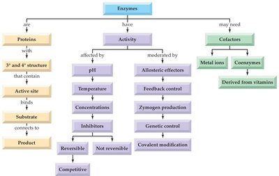
Enzymes in metabolic pathways are regulated to ensure proper cellular function. Regulation can be achieved through several mechanisms:
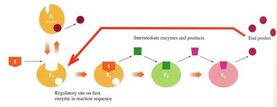
Feedback Control: The end product of a pathway acts as an allosteric regulator of the first enzyme, either inhibiting (negative feedback) or enhancing (positive feedback) its activity.
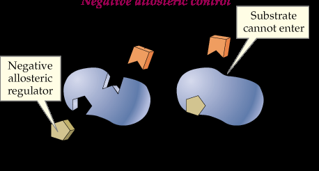
Allosteric Regulation: Molecules bind to sites other than the active site, causing conformational changes that affect enzyme activity. Positive regulators increase activity, while negative regulators decrease it.
Covalent Modification: Addition or removal of functional groups (e.g., phosphorylation) alters enzyme activity.
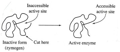
Zymogen Activation: Enzymes are synthesized as inactive precursors (zymogens) and activated by removal of a protein segment.
Genetic Control: Regulation of enzyme levels through gene expression.


Vitamins and Coenzymes
Classification of Vitamins
Vitamins are essential organic molecules required for various biological functions. They are classified based on their solubility:
Water-Soluble Vitamins: Hydrophilic, contain OH and COOH groups, not stored in the body, often function as coenzymes. Examples include Vitamin C and B-complex vitamins.
Fat-Soluble Vitamins: Hydrophobic, composed mainly of carbon and hydrogen, stored in the liver. Examples include Vitamins A, D, E, and K.
Reason for Classification: Solubility affects storage, absorption, and excretion. Water-soluble vitamins are excreted in urine, while fat-soluble vitamins can accumulate in tissues.
Fat-Soluble Vitamins
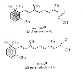
Vitamin A (Retinol): Provitamin: β-carotene. Functions in vision, cell growth, and as an antioxidant. Deficiency leads to blindness and growth issues; toxicity causes liver damage.
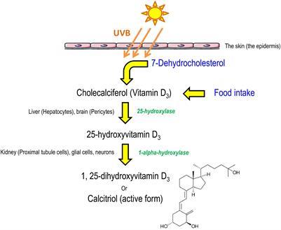
Vitamin D (Calciferol): Provitamin: 7-dehydrocholesterol. Required for bone growth and calcium absorption. Deficiency causes rickets; toxicity leads to calcification of tissues.
Vitamin E (α-tocopherol): Strong antioxidant, prevents oxidative damage. Deficiency is rare; toxicity interferes with blood clotting.
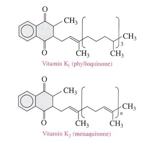
Vitamin K (Phylloquinone, Menaquinone): Required for blood clotting and bone metabolism. Deficiency causes bleeding disorders; toxicity leads to liver dysfunction.
Water-Soluble Vitamins and Coenzymes
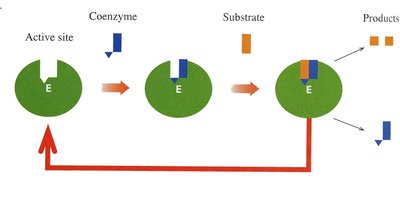
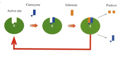
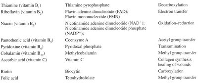
Water-soluble vitamins often function as coenzymes, which are organic compounds required for enzyme catalysis. Most coenzymes are derivatives of B vitamins.
Vitamin B1 (Thiamine): Coenzyme: Thiamine pyrophosphate (TPP). Function: Decarboxylation in carbohydrate metabolism.
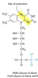
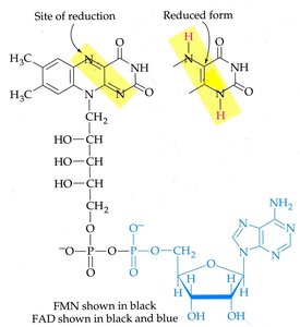
Vitamin B2 (Riboflavin): Coenzymes: FMN, FAD. Function: Redox reactions.
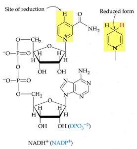
Vitamin B3 (Niacin): Coenzymes: NAD+, NADP+. Function: Redox reactions.
Vitamin B5 (Pantothenic Acid): Coenzyme: Coenzyme A. Function: Acetyl group transfer.
Vitamin B6 (Pyridoxine): Coenzyme: Pyridoxal phosphate (PLP). Function: Amino acid metabolism.
Vitamin B7 (Biotin): Coenzyme: Biotin. Function: Carboxylation reactions.
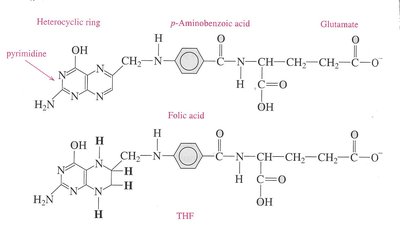
Vitamin B9 (Folic Acid): Coenzyme: Tetrahydrofolate (THF). Function: One-carbon transfer in nucleic acid synthesis.
Vitamin B12 (Cobalamin): Coenzyme: Methylcobalamin. Function: Methyl transfer and nucleic acid metabolism.
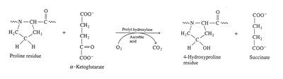
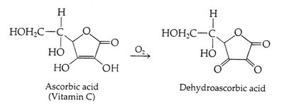
Vitamin C (Ascorbic Acid): Function: Antioxidant, collagen synthesis, healing wounds.
Diagnostic Enzymes and Serological Testing
Enzyme Markers for Disease Diagnosis
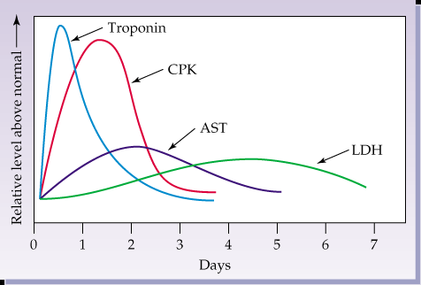
Enzyme activity in blood can be used to diagnose organ damage or disease. Specific enzymes are associated with particular tissues:
Liver: AST, ALT, GGT, ALP
Heart: CK (CPK), LDH, Troponin
Pancreas: α-amylase
Prostate: PSA, Acid Phosphatase
Isozymes: Different quaternary forms of the same enzyme, tissue-specific, useful for pinpointing organ damage.
Serological Testing and Lateral Flow Assays
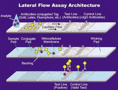
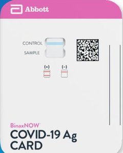
Serological tests, such as lateral flow assays, are used for rapid detection of antigens (e.g., COVID-19). These tests utilize protective proteins called antibodies to bind specific antigens.
Lateral Flow Assay: Uses antibodies conjugated to tags to detect the presence of viral proteins (e.g., SARS-CoV-2 spike protein).
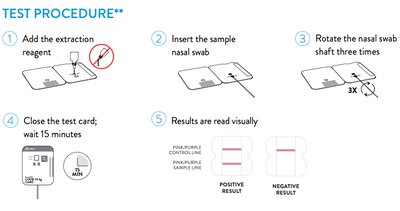
Test Procedure: Sample is applied, flows through the membrane, and results are read visually.
Summary Table: Water-Soluble Vitamins and Coenzymes
Vitamin | Coenzyme | Function |
|---|---|---|
Thiamine (B1) | Thiamine pyrophosphate | Decarboxylation |
Riboflavin (B2) | FAD, FMN | Electron transfer |
Niacin (B3) | NAD+, NADP+ | Oxidation-reduction |
Pantothenic acid (B5) | Coenzyme A | Acetyl group transfer |
Pyridoxine (B6) | Pyridoxal phosphate | Transamination |
Cobalamin (B12) | Methylcobalamin | Methyl group transfer |
Ascorbic acid (C) | Vitamin C | Collagen synthesis, healing wounds |
Biotin (B7) | Biocytin | Carboxylation |
Folic acid (B9) | Tetrahydrofolate | Methyl group transfer |
Summary Table: Fat-Soluble Vitamins
Vitamin | Provitamin | Functions |
|---|---|---|
Vitamin A (retinol) | β-carotene | Vision, cell growth, antioxidant for lipids |
Vitamin D (calciferol) | 7-dehydrocholesterol | Bone health, calcium and phosphate absorption |
Vitamin E (α-tocopherol) | None | Antioxidant, prevents oxidative damage |
Vitamin K (phylloquinone) | None | Blood clotting, bone metabolism |
Additional info: These notes expand on the original lecture content by providing definitions, examples, and summary tables for clarity and completeness. All images included are directly relevant to the adjacent explanations.
