 뒤로
lec 9 Protein Structure and Function: GOB Chemistry Study Notes
스터디 가이드 - 스마트 노트
 자료에 맞춘 맞춤형 노트, 핵심 정의, 예시, 맥락을 확장해 제공합니다.
Protein Structure and Function
Levels of Protein Structure
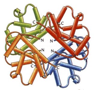
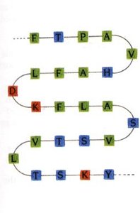
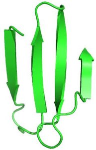
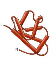
Proteins are complex biomolecules whose structure determines their function. The four levels of protein structure are primary, secondary, tertiary, and quaternary, each contributing to the overall shape and activity of the protein.
Primary Structure: The sequence of amino acids in a polypeptide chain, held together by peptide bonds.
Secondary Structure: Local folding patterns such as alpha helices and beta sheets, stabilized by hydrogen bonds.
Tertiary Structure: The three-dimensional shape of a single polypeptide chain, stabilized by interactions among side chains.
Quaternary Structure: The arrangement of multiple polypeptide chains (subunits) into a functional protein complex.
Shape Determining Interactions
The folding and stability of proteins are determined by covalent and noncovalent interactions:
Covalent Bonds: Include peptide bonds and disulfide bonds (S-S) between cysteine residues.
Electrostatic Interactions: Salt bridges formed between acidic and basic side chains.
Hydrogen Bonds: Occur between H-bond donors and acceptors (O-H, N-H).
Van der Waals Forces: Weak interactions between all atoms.
Classification of Protein Structures
Proteins are classified based on their shape and solubility:
Globular Proteins: Globe-like, typically water soluble, with hydrophilic residues on the surface. Examples include enzymes, myoglobin, and hemoglobin.
Fibrous Proteins: Form fibers or sheets, typically insoluble, with tightly packed hydrophobic residues. Examples include collagen, keratin (hair, teeth, fingernails).
Conjugated Proteins
Types of Conjugated Proteins
Conjugated proteins require non-protein components for function. The main types are:
Glycoproteins: Covalently bound carbohydrates (e.g., cell surface receptors).
Lipoproteins: Covalently bound lipids (e.g., cholesterol transporters).
Metalloproteins: Contain metal ions for structure or catalytic function (e.g., zinc-stabilized proteins).
Phosphoproteins: Covalently bound phosphate groups for activation/deactivation or binding sites.
Hemoproteins: Contain heme groups/porphyrin rings (e.g., oxygen transporters).
Nucleoproteins: Contain nucleic acids (e.g., ribosomes).
Myoglobin and Hemoglobin
Structure and Function
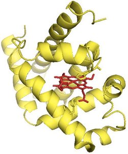
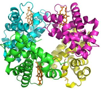
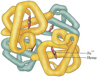
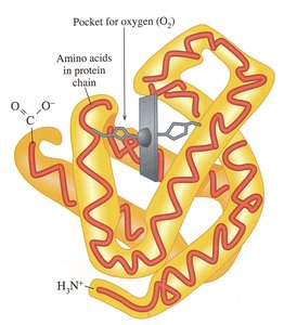
Myoglobin and hemoglobin are globular conjugated proteins involved in oxygen transport and storage.
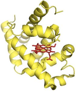
Myoglobin: Found in muscles, consists of a single polypeptide chain (tertiary structure), stores oxygen.
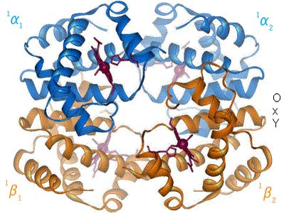
Hemoglobin: Found in red blood cells, consists of four polypeptide chains (quaternary structure), transports oxygen.
Oxygen Binding and Transfer
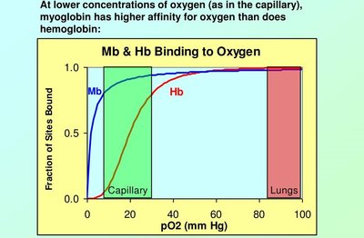
Hemoglobin undergoes conformational changes upon oxygen binding, facilitating oxygen transfer to myoglobin in muscle tissues.
Oxygen Affinity: Myoglobin has higher affinity for oxygen at low concentrations, hemoglobin releases oxygen in capillaries.
Conformational Change: Oxygen binding causes structural changes in hemoglobin's quaternary structure.
Sickle Cell Anemia
Structural Changes and Disease Mechanism
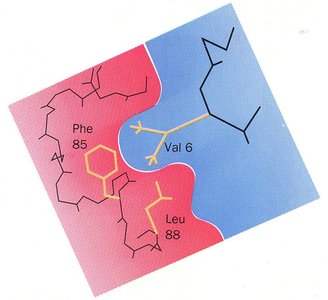
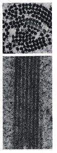
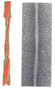
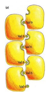
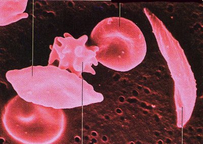
Sickle cell anemia is caused by a mutation in hemoglobin, where glutamic acid is replaced by valine at position 6, creating a hydrophobic patch that leads to aggregation and fibrous structure formation.
Mutation: Glu → Val at position 6 in the beta chain.
Aggregation: Hydrophobic valine interacts with hydrophobic pockets on adjacent molecules, causing aggregation.
Symptoms: Anemia, pain, stroke, fatigue; sickled RBCs have reduced lifespan.
Electrophoresis: Analytical and Diagnostic Tool
Principle and Application
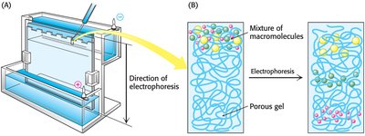
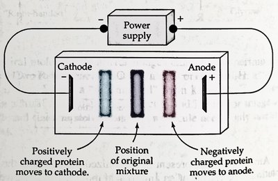
Electrophoresis separates proteins based on charge and molecular weight, useful for diagnosing diseases such as sickle cell anemia.
Charge Separation: Proteins migrate towards anode or cathode depending on their charge.
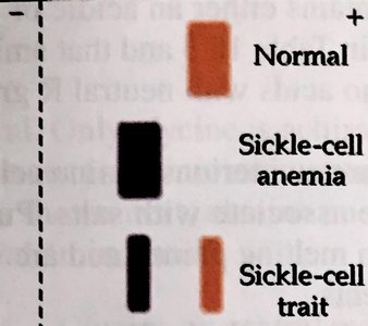
Diagnostic Use: Sickle cell hemoglobin migrates differently than normal hemoglobin due to the mutation.
Collagen: Structure and Related Diseases
Structure and Function
Collagen is a fibrous protein with quaternary structure, providing strength and support to connective tissues, bones, and teeth.
Subunit: Tropocollagen, stabilized by hydrogen bonds.
Amino Acid Composition: Every third residue is glycine for close packing; proline and hydroxyproline are also abundant.
Stabilization: Covalent (disulfide) bonds between strands.
Collagen Related Diseases
Brittle Bone Disease (Osteogenesis imperfecta): Insufficient or poor quality collagen, leading to fractures and weak tissues.
Scurvy: Vitamin C deficiency impairs collagen synthesis, causing skin lesions, joint pain, and gum disease.
Keratin: Structure and Stabilization
Alpha and Beta Keratin
Keratin is a fibrous protein found in hair, nails, and skin. Its structure is stabilized by disulfide bonds (S-S), which provide strength and resilience.
Alpha-Keratin: Stabilized by disulfide bonds between peptide chains.
Beta-Keratin: Stabilized by van der Waals interactions.
Protein Folding and Chaperones
Chaperones and Protein Folding
Chaperones are proteins and protein complexes that assist in the folding of other proteins, preventing aggregation and ensuring proper function.
Function: Prevent misfolding and aggregation of proteins.
Importance: Not all proteins fold correctly during synthesis; chaperones are essential for cellular health.
Prions: Misfolded Infectious Proteins
Prion Diseases
Prions are misfolded proteins in the central nervous system that induce other proteins to misfold, causing transmissible spongiform encephalopathies (TSEs), which are fatal degenerative brain disorders.
Mechanism: Conversion of alpha-helices to beta-sheets, inducing misfolding in other prions.
Transmission: Through infected tissue, fluids, or contaminated instruments; resistant to normal sterilization.
Protein Degradation and Denaturation
Protein Degradation
Protein degradation is the breakdown of proteins into amino acids, occurring via hydrolysis of peptide bonds. It is essential for removing misfolded or unneeded proteins and recycling cellular components.
Autophagy: Self-degradative process for energy balance and removal of damaged components.
Lysosomes: Cellular organelles that degrade unwanted material.
Proteasomes: Degrade unfolded or unneeded proteins by proteolysis.
Protein Denaturation
Protein denaturation is the unfolding of proteins, disrupting their structure and function. It is primarily a physical process caused by various agents:
Heat: Disrupts non-covalent interactions.
Mechanical Agitation: Causes foaming and loss of water.
Detergents: Interact with hydrophobic side chains.
Organic Compounds: Disrupt hydrogen bonds (e.g., ethanol).
pH Change: Alters charge, disrupting salt bridges.
Inorganic Salts: Disrupt salt bridges at high concentrations.
Summary Table: Types of Protein Structure
Structure Level | Description | Stabilizing Forces |
|---|---|---|
Primary | Amino acid sequence | Peptide bonds |
Secondary | Alpha helices, beta sheets | Hydrogen bonds |
Tertiary | 3D folding of single chain | Hydrogen bonds, ionic bonds, disulfide bonds, van der Waals |
Quaternary | Assembly of multiple chains | Same as tertiary, plus inter-chain interactions |
Key Equations
Peptide Bond Formation:
Protein Hydrolysis:
Additional info: These notes expand on the lecture content with definitions, examples, and context suitable for GOB Chemistry students, covering protein structure, function, disease relevance, and analytical techniques.
